

 PDF
PDF Citation
 Print
Print


INTRODUCTION
Pemphigus diseases are a group of autoimmune disorders that have certain common features, and these diseases are considered to be potentially fatal1,2. Pemphigus vegetans is a variant of pemphigus vulgaris and is the rarest form of pemphigus; Pemphigus vegetans comprises less than 1~2% of all pemphigus cases1,3,4. This variant is characterized by flaccid bullae or pustules that erode to form hypertrophic papillated plaques that predominantly involve the intertriginous areas, the scalp, and the face; in 60~80% of all cases, the oral mucosa are also affected5,6. Clinically, two subtypes are recognized: the Neumann and Hallopeau subtypes1,3. The Neumann type is characterized by bullae that extend and coalesce; they then evolve to vegetating masses which become dry, hyperkeratotic, and fissured. The Hallopeau type is characterized by a polycyclic eruption of pustules that form firm pink papillomas which progressively flatten and change to dark brown plaques with a benign course and few relapses4,7,8.
We report here on a 63-year-old woman with a Neumman type of pemphigus vegetans who was was successfully treated with dapsone.
CASE REPORT
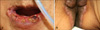
A 63-year-old woman presented with a 2-year history of vegetating, papillomatous plaques on the inguinal folds and erosions of the oral mucosa, tongue, and perioral area. The cutaneous lesions started with vesicles and bullae that extended peripherally and later formed the vegetating lesions. No other lesions on the skin were seen, and there was no history of other skin disease. She had no family history of a blistering disorder. On physical examination, there were oozing, erosive vesicles on the lip and hypertrophic verrucous vegetative plaques on the inguinal folds (Fig. 1). Laboratory assessments showed an absolute eosinophil count of 20.7% (normal range: 0~5%) and a lymphocyte count of 17.6% (normal range: 20~44%). The results of routine serum chemistry, including liver function tests, were within the normal ranges.
Biopsy specimens were obtained from the erosive vesicles on the lip and a verrucous plaque on the inguinal folds. The histologic findings of the erosive vesicles from the lip revealed a suprabasal cleft with scattered acantholytic cells (Fig. 2A). Biopsy of the vegetating plaque from the inguinal folds showed massive papillomatosis and spongiosis, as well as eosinophilic granulocytes throughout the entire thickness of the epithelium. In addition to eosinophilic spongiosis, we also found sharply bordered, eosinophilic abscesses in all the layers of the epidermis. Eosinophils also dominated the dense inflammatory infiltrate in the papillary dermis (Fig. 2B). Antibodies to desmoglein 1 and 3 were analyzed using a specific enzyme-linked immunosorbent assay (ELISA). The results of ELISA with recombinant purified desmoglein (Dsg) 1 and Dsg3 (Medical & Biological Laboratories corporation, Nagoya, Japan) were positive for Dsg3 (a titer of 172.2 U/ml) but not Dsg 1. Based on the clinical, histopathologic, and ELISA findings, pemphigus vegetans as a definite diagnosis was made.
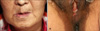
The patient was treated with oral methylprednisolone 16 mg daily, but no improvement was observed after 7 days of treatment. Subsequently, treatment with a combination of oral methylprednisolone 12 mg daily and dapsone 50 mg daily was started. The erosive vesicular and verrrucous lesions were healed 3 weeks after this combination therapy (Fig. 3). During treatment, the eosinophil count returned to normal. The doses of methylprednisolone were tapered to 8 mg daily without the appearance of new lesions. The patient is presently well controlled on maintenance therapy of 4 mg methlyprednisolone and 50 mg dapsone daily. She has not had any recurrence of the lesions since.
DISCUSSION
Pemphigus vegetans is a rare clinical form of pemphigus and is characterized by verrucous intertriginous plaques1,2. Analysis of the demographic and clinical data of the reported cases of pemphigus vegetans in the literature reveals that individuals of all ages, including children, can be affected by the disease9,10.
Pemphigus vegetans is classified based on the initial clinical picture and disease course as the Neumman type or Hallopeau type2,7. Consistent with Neumann type disease, our patient had initial mucosal erosions and disseminated, slightly rupturing blisters on the intertriginous areas, which formed the basis for papillomatous vegetations4,5. Oral involvement is present in nearly all pemphigus vegetans cases. A characteristic feature of pemphigus vegetans is the cerebriform tongue, which is characterized by a pattern of sulci and gyri on the dorsum of the tongue9,10.
Pemphigus vegetans is caused by intercellular autoantibodies primarily against desmogleins 1 and 3, which are adhesion molecules in the desmosomes of keratinocytes1,2,4. Previous studies have consistently reported autoantibodies against desmoglein 3 in patients with pemphigus vegetans, and our patient was no exception. Autoantibodies against desmoglein 1, desmocollin 1 and 2, and periplakin have occasionally been detected in previously reported cases4,11,12. Captopril has been reported to induce this disease9,13. Development of pemphigus vegetans in intranasal heroin users has also been reported, but this relationship remains uncertain13,14.
Histopathologically, the early lesions of pemphigus vulgaris and pemphigus vegetans show suprabasal acantholysis1,3. Pemphigus vegetans also exhibits epidermal hyperplasia, papillomatosis, and intraepidermal eosinophilic abscesses as the lesions age. The histopathologic changes in pemphigus vegetans seem to differ from those of the vulgaris type by an eosinophic response, formation of microabscesses, and the extent of vesiculation3,4. The immunofluorescence findings in pemphigus vegetans are indistinguishable from those of pemphigus vulgaris3,11. Direct immunofluorescence demonstrates deposition of IgG and C3 on the cell surface of keratinocytes. Indirect immunofluorescence reveals circulating antiepithelial cell-surface IgG4,11. However, our patient refused skin biopsy for immunofluorescence because of her economic status, so an immunofluorescence study was not performed.
It might be difficult to make a diagnosis precisely due to the varied clinical presentation and the histopathological resemblance to other conditions. Some diseases or disorders have to be differentiated from pemphigus vegetans1,2. These include the vegetating lesions of other bullous autoimmune skin diseases, such as bullous pemphigoid or IgA pemphigus, the chronic inflammatory plaques in Haiely-Haiely disease, and especially vegetating pyoderma1,3. Making the differential diagnosis from pyostomatitis vegetans is especially important. Both conditions show similar clinical and histopathological findings, and only direct and indirect immunofluorescence studies enable the clinician to distinguish between these two disorders. Only the lesions of pemphigus vegetans characteristically show epithelial intercellular deposits of immunoglobulins3,15. Our patient had antibodies directed against demoglein 3, and the final diagnosis of pemphigus vegetans was made.
Treatment of pemphigus vegetans is similar to that for pemphigus vulgaris, and this is normally accomplished with systemic steroids1,16. However, oral administration of corticosteroids can not always induce disease remission. The addition of immunosuppressants, such as azathioprine, mycophenolate mofetil, or cyclosporine may improve remission rates and allow a steroid-sparing effect16,17. Patients with the Neumann type have a similar course as those with pemphigus vulgaris; they require higher doses of corticosteroids and have relapses and remissions. Hallopeau patients have fewer relapses if any, and they usually respond to lower doses of corticosteroids. Studies on on the successful use of dapsone and retinoids have also been published8,16,18.
In a study by Gürcan and Ahmed, 32 patients with pemphigus vulgaris and 14 patients with pemphigus foliaceus responded to dapsone19. The overall response rates to dapsone, when given either alone or in combination with corticosteroids or immunosuppressive agents, were 84% for mucous membrane pemphigoid and 81% for bullous pemphigoid. The adverse effects of dapsone are dose-dependent and are usually reversible19.
One study showed that dapsone inhibits neutrophil adherence to pathogenic IgG in bullous pemphigoid and to IgA in patients with IgA dermatoses in a dose-dependent manner17,19. Dapsone also inhibits the release of bullous pemphigoid IgG-induced interleukin-8 from cultured normal human epidermal keratinocytes by mechanisms that act at the post-transcriptional level19,20. An additional mechanism that may account for the effectiveness of dapsone during the maintenance phase is inhibition of eosinophilic spongiosis. Dapsone has been shown to be an inhibitor of eosinophilic peroxidase, which is a mediator of inflammation that may play a role in the spongiotic events observed in pemphigus lesions17,20.
The treatment of pemphigus disorder with dapsone is less expensive than the immunomodulatory agents that are often administered. Dapsone reduces the steroid dose in patients with autoimmune mucocutaneous blistering diseases; thus, it can be used as a corticosteroid-sparing agent. Therefore, its combination use with oral corticosteroids may be useful in pemphigus disorders. Thus, we propose that dapsone might be an effective adjunct in the treatment for patients with pemphigus vegetans.

 XML Download