

 PDF
PDF Citation
 Print
Print


INTRODUCTION
Calcium channel blockers (CCB) are rarely associated with adverse skin reactions; the incidence of reactions is 8 per 106. prescriptions for nifedipine and diltiazem, with reactions consisting mainly of pruritus and urticaria1. Only seven cases of telangiectasia appearing in photo distributed areas as a result of administration of CCB have been reported1-5, three of these cases were induced by amlodipine2,3,5.
CASE REPORT
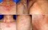
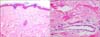
A 56-year-old man presented with a two-year history of non-pruritic telangiectasia, which initially developed on the face and spread to the neck and both shoulders. His hypertension was treated with amlodipine for two years without any additional medications. He had no history of photosensitivity, rosacea, or use of topical steroids. Physical examination revealed asymptomatic widely distributed spider-like telangiectasia on the face, V area of the neck, and the shoulders (Fig. 1A). Biopsy specimens taken from his arm revealed enlarged capillaries in the upper dermis without signs of vasculitis (Fig. 2). Laboratory studies revealed a normal total white cell count and liver enzymes. Due to his clear history and time consistencies between onset of the cutaneous reaction and administration of amlodipine, a diagnosis of photodistributed amlodipine induced-telangiectasia was made. Amlodipine was discontinued and replaced by irbesartan and thiazide. A remarkable improvement was observed 1 month later and two months later; the telangiectasia had almost faded. However, after 4 months, he was admitted to the hospital with cerebral infarct and uncontrolled hypertension. Amlodipine was re-administrated and the rash relapsed within 1 week (Fig. 1B). He has been receiving maintenance therapy with clopidogrel and amlodipine with persistent telangiectasia on the face and shoulders.
DISCUSSION
Iatrogenic telangiectasia is a poorly understood dermatological side-effect, occurring secondary to administration of many drugs, including lithium, thiotrixene, interferonalfa, and isotretinoin6. Telangiectasia, localized to photoexposed sites, has been described after administration of calcium channel blockers1-5. In addition, two cases of photo-distributed telangiectasia resulting from cefotaxime and venlafaxine have been reported6,7.
CCB is the first-line therapy in treatment of hypertension. The three subclasses of CCB include the following: verapamil, diltiazem, anddihydropyridines (amlodipine, felodipines, and nifedipine). Almost all reported cases of calcium
channel blocker-induced telangiectasia were from dihydropyridines, due to their greater vascular smooth muscle effects relative to cardiac effects, compared with diltiazem and verapamil8). CCB may provoke telangiectasia by the nature of their vasodilatory action; however, it is not clearly understood how they may cause photosensitivity. One theory is that the photoproducts created by UVA and visible wavelengths bind with skin proteins, leading to telangiectasia2. This cutaneous reaction is regarded as photosensitive; however, it does not clearly correspond to the signs and symptoms of a photoallergic or phototoxic reaction.
Differential diagnosis of telangiectasia considers many underlying diseases, including liver disease, essential telangiectasia, scleroderma, and lupus erythematosus5; the majority of these can be excluded by a careful history, examination, and skin biopsy.
In summary, the photo-distributed telangiectasia observed in our patient, together with the considerable improvement seen within two months of stopping amlodipine and recurrence after restarting, is clearly suggestive that amlodipine was the causative drug. It is important to consider that use of these drugs can result in acquired telangiectasia and prompt discontinuation may lead to spontaneous improvement. However, close observation and monitoring of blood pressure after changing these drugs should be required.
 XML Download