

 PDF
PDF Citation
 Print
Print


INTRODUCTION
Pincer nail deformity has been characterized by the excessive curvature of the nail plate in the transverse dimension. Three main forms of this overcurvature have been identified: an arched nail, a tile-shaped nail, and a plicated nail1. This overcurvature has been shown to increase from the proximal to the distal direction, leading to pinching off and loss of soft tissue of the involved digits, causing severe pain2. This deformity has been attributed to both hereditary and acquired causes, including psoriasis, trauma, developmental abnormalities, treatment with beta blockers, allergic reactions, epidermal cysts, subungual exostosis, osteoarthritis, and ill-fitting shoes3. Numerous treatment modalities have been described for correcting this nail deformity, including both conservative and surgical methods4-9. The conservative approaches have entailed a long treatment duration and the corrective results not always have been permanent. Further, most surgical methods also have been unsatisfactory in terms of relapse and cosmetic outcome. Therefore, dermal grafting was employed in the correction of a plicated nail deformity in the left thumb of a 49-year-old woman with favorable results.
CASE REPORT
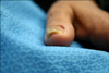
A 49-year-old female presented with a nail deformity of the left thumb which had been present for many years. With the increasing severity of the deformity, the patient had been experiencing increased pain severity. The patient was otherwise healthy, and had no family history of a similar lesion. Upon physical examination, a dystrophic left thumb nail was noted with prominent right axial curvature that caused compression of the distal portion of the thumb (Fig. 1). Radiographic examination showed no evidence of osteophyte formation at the base of the distal phalanx, and fungal nail infection was excluded by negative results of microscopic examination (KOH test) and fungal culture.
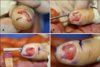
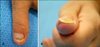
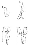
Initially, two percent lidocaine without epinephrine was administered via the standard technique to achieve digital block and wing block at the left thumb. The thumb was wrapped with a surgical glove and the glove-finger was released from the finger tip down to the proximal part of the finger and fixed by a mosquito clamp to provide a finger tourniquet. The deformed nail plate was removed from the underlying nail bed by the rolling method. An oblique incision was made distal to the hyponychium in line with the most lateral aspect of the nail bed (Fig. 2A), and a tunnel for the graft was created between the nail bed and the underlying phalanx with a periosteal elevator (Fig. 2B). The dermal graft was harvested under local anesthesia from the thenar area of the left hand. The graft length ranged from the hyponychium to the paronychial fold and was at least one centimeter in width. The epidermis and subcutaneous fat were removed by fine scissors, and the donor site was finalized by primary closure. The graft was put into the hyponychial incision and drawn proximally in the tunnel more than one centimeter away from the nail matrix by passing the graft (distal to proximal) through the tunnel by nylon 4-0 sutured to the distal end of graft without incising the paronychial nail fold (Fig. 2C). When the graft elevated the paronychial fold, it sutured the skin proximally to hold it within the tunnel. The excess portion of the graft was excised distally and the hyponychial incision was closed with nylon 4-0 (Fig. 2D). Fig. 3 are post-surgical results of the patient after 6 weeks, 5 months respectively. The patient has been doing well, and there was no relapse since in the 12 months following the procedure. Schematic surgical procedures have been provided in Fig. 4.
DISCUSSION
The progressive tubulization of a nail plate not only affects its cosmetic appearance, but also causes pain, leading to substantial discomfort in daily life. Further worsening of the curvature results in pinching of the subungual tissue. The pathogenesis of acquired pincer nail deformity has been attributed to many factors, including psoriasis, trauma, developmental abnormalities, administration of beta blockers, allergic reactions, epidermal cysts, subungual exostosis, osteoarthritis, and ill-fitting shoes3. Cases in which the deformity was inherited have often showed changes similar to those seen in other family members. Previous studies have reported evidence for an autosomal-recessive inheritance pattern10, however, no such contributing factors were found in our case.
Several methods have been reported for the treatment of pincer nail deformity, including both conservative methods such as the use of uric acid, clipping, the attachment of plastic braces, and splint fixation, and surgical methods such as nail plate removal and nail bed ablation4-8, depending on the degree of severity7,11. However, most of the methods have been unsatisfactory in terms of recurrence and cosmetic outcome.
Recent surgical methods used in the Korean dermatologic field were introduced by Moon et al.4 and Yun et al.12. According to them, no recurrence occurred during a follow-up period of 14 to 24 months and 18 months, respectively4,12. However, if the curvature of the nail plate differs between left and right sides, it is possible that the nail plate newly formed after treatment may also show axis deviation. Furthermore, cases with different curvature have been much more common than those with the same curvature on the left and right sides of nail plate.
In our patient, the nail plate curvature of the thumb was different on both sides, prominently curved on the right side. For these reasons, we decided to use the dermal grafting method to correct the pincer nail deformity. Restoration of the nail bed contour by surgically implanting a dermal graft under the affected nail bed resulted in long-term correction while preserving the nail matrix13. In our patient, follow-up examination showed correction of the excessive nail curvature, which previously caused pain and swelling around the nail plate.
The dermal grafting method has been already described elsewhere3,13, and the results were reported to be very good in all. Although this method has not been popular in the dermatologic field and although it has many disadvantages including several technical difficulties and a long operative time, we think this is a very effective method and has the advantages of relatively low recurrence rate and being successful even in cases of pincer nails showing different nail plate curvature on both sides.
 XML Download