

 PDF
PDF Citation
 Print
Print


INTRODUCTION
Pseudocyst of the scalp was first described in 1992 by Iwata et al.1 in a report of 19 Japanese patients presenting with alopecia and a solitary subcutaneous tumor with a histologically ill-defined cyst wall. In 1998 in France, Chevallier2 reported 3 cases described as 'noninfectious abscess of the scalp with alopecia'. In 2011, Abdennader et al.3 reported 15 cases with similar morphological features to those of pseudocyst of the scalp; however, the condition was referred to as "alopecic and aseptic nodules of the scalp" because pseudocyst formation was not always present. Here, we report a case of pseudocyst of the scalp and summarize the characteristic features of both pseudocyst of the scalp and alopecic and aseptic nodules of the scalp.
CASE REPORT
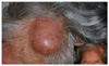
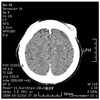
A 72-year-old woman presented with a 2-month history of a localized fluctuating mass on her scalp. On examination, a skin-colored, dome-shaped cystic tumor measuring 50 mm in diameter was noted with alopecia in the same area as the lesion (Fig. 1). The patient did not complain of symptoms such as itching, pain, or tenderness inthelesion. Multi-detector computed tomography suggested that the lesion was not fixed to the bone and was limited to the skin above the skull (Fig. 2). The patient had no history of an insect bite, trauma, or medication. Her medical history included herpes zoster along the V1 dermatome of the left scalp that had been successfully treated with an oral antiviral agent 5 years earlier. Laboratory tests showed no remarkable findings.
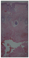
Accordingly, the lesion was surgically resected under local anesthesia. The puncture material was bloody and sterile and produced negative bacteriological and mycological culture results. Microscopic examination of the biopsy showed a pseudocyst-like architecture and fibrosis in the lower dermis (Fig. 3). There was no evidence of a true cystic wall. The skin lesion was treated with total excision, sutures, and a pressure dressing of packing gauze for 5 days. Five months after treatment, the skin lesion had not recurred and the patient was left with an excellent cosmetic result.
The clinical history and histopathological findings described here resemble those for pseudocyst of the scalp1.
DISCUSSION
Pseudocyst of the scalp was first described in the Japanese literature by Iwata et al. in 19921. They reported 19 cases of a dome-shaped subcutaneous tumor with alopecia and an ill-defined cyst wall. Such tumors, which are seen predominantly in men aged between 18 and 40 years, are located between the top and forehead area of the scalp. Subsequently, several reports of similar cases have appeared in the Japanese literature4-7.
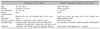
In 1998, Chevallier2 reported three cases of "noninfectious abscess of the scalp with alopecia" that were similar in morphology to pseudocyst of the scalp. Additionally, Abdennader et al.3 reported 15 cases with similar features to those of pseudocyst of the scalp, which were referred to as alopecic and aseptic nodules of the scalp (AANS). Since then, the condition has been characterized as largely asymptomatic tumors located mainly at the vertex and upper portion of the occipital area, with most patients being men between the age of 12 and 38 years2. The characteristic findings in pseudocyst of the scalp and AANS are similar except for those revealed by histology and from puncture material (Table 1).
The etiology is unknown, but the pseudocyst is considered to initially develop as folliculitis associated with follicular occlusion1 or foreign body2. When follicular occlusion occurs, a granulomatous infiltrate is generated accompanied by acute inflammation with histiocytes, lymphocytes, and giant cells clustered around each follicle. Subsequently, the central tissue undergoes necrosis and blood and lymphatic vessels are broken, resulting in a bloody, yellowish exudate and pseudocyst formation. The etiology of AANS is also unknown. It is probably a particular form of deep folliculitis leading to a non-scarring type of alopecia. It might represent a part of a spectrum including several diseases such as acne conglobata and hidradenitis suppurativa, in which a follicular occlusion is suspected as the cause of the cyst or pseudocyst. Further studies are obviously needed to clarify the pathogenesis of AANS3.
The histopathology of pseudocyst of the scalp shows a pseudocyst and granuloma in deep dermis1. However, no granuloma was found in our case. Racial factors where the patient has a different type of hair or there is a different period of the cyst development might explain these findings.
The differential diagnosis of pseudocyst of the scalp includes a ruptured epidermal cyst1 and dissecting cellulitis of the scalp8. Ruptured epidermal cysts are soft with a central blackish dot, and are not associated with alopecia. Furthermore, an epidermal lining with a granular layer or keratin is not found in pseudocyst of the scalp. Dissecting cellulitis of the scalp is characterized by sinus tracts with scarring alopecia.
The successful treatment of pseudocyst of the scalp will resolve the lesions without subsequent recurrence and alopecia. Several treatments have been reported, including needle aspiration, surgical excision, steroidal injection, and the administration of doxycycline at 100 mg/day1,4-7. The patient described here was treated by surgical excision and pressure dressing; at the time of writing, the patient's skin lesions had not recurred. In AANS, the treatment with doxycycline is efficient at the dose of 100 mg/day. However, the duration of the treatment must be at least 3 months3.
In summary, we reported a case of pseudocyst of the scalp in Korea. More work must be conducted to elucidate the pathogenesis of this disease entity and to establish the most effective method of treatment.
 XML Download