

 PDF
PDF Citation
 Print
Print


INTRODUCTION
A petrified auricle is an unusual clinical entity in which the ear becomes partially or completely rigid. This may result from frostbite, local trauma, inflammation, or systemic diseases1. Ossification of the auricle is a rare condition caused by the petrification of the auricle.
In 1866, a Polish anatomist Bochdalek2 described a case of auricular ossification with histopathological confirmation. Later, in 1899 Wassmund3 reported X-ray findings of auricular ossification in a patient.
Here, we describe a case of a man who developed ossification of the left ear after repeated episodes of irritation. Ossification of skin can be clinically considered as a relatively common condition; however, reports on auricular ossification are rare in Korea.
CASE REPORT
A 53-year-old man developed gradual rigidity and thickening of the left auricular cartilage 1 year ago. He works in a steel factory and has a 2-year history of hearing loss in the left ear because of a cochlear defect. He had heard that stimulation of the auricle was a good method to recover the auditory sense. Since then, he has been rubbing his left auricle repeatedly. He did not have any history of participating in boxing or wrestling, and had no history of any other physical trauma or ear inflammation. His further medical history was unremarkable, and he did not use any regular medication.
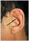
Clinical examination of the left ear revealed that the auricle was rigid and had decreased flexibility (Fig. 1). Moreover, thickening of the skin (lichenification) was observed as a result of chronic rubbing. The earlobe had a normal configuration and did not show any sign of epidermal destruction. Auricular tactile sensation was appropriate. The right ear was not affected.
Radiographs demonstrated indistinct ossification of the auricular cartilage. Histopathological examination revealed that the epidermis was hyperkeratosis and irregular acanthosis, and that the elastic cartilage was being replaced by a lamellar bone due to calcium deposition (Fig. 2).
Additional laboratory tests, including complete blood cell count, serum calcium, serum phosphorus, serum alkaline phosphatase, glucose, electrolytes, thyroid function tests, and parathyroid hormone tests showed findings within the normal range.
Thus, ossification of the left auricle was diagnosed based on these clinical, radiographic, and histological results. Our patient's complaint was only of mild discomfort when sleeping, thus he refused treatment. He was advised to stop inducing any mechanical skin irritation on his left auricle.
DISCUSSION
Cartilage is classified into elastic, hyaline and fibrocartilage, based on the presence of elastic fibers and collagen and on tissue morphology. Elastic cartilage is a basic component of the auricle, external ear canal, nose, and epiglottis in the head and neck and does not normally ossify or calcify. Based on the absence or presence of previous cutaneous lesions, cutaneous ossification is classified as primary or secondary, respectively. Primary ectopic ossification is seen in rare syndromes, including Albright's hereditary osteodystrophy, fibrodysplasia ossificans progressiva, and osseous heteroplasia4. Ectopic auricular ossification is a type of secondary cutaneous ossification that generally occurs due to trauma or in certain neoplastic processes, and is often preceded by calcification5,6.
The occurrence of auricular ossification is rare and is more frequently observed in men than in women. Although unilateral manifestation has been reported, bilateral auricular ossification apparently occurs more often7. The clinical presentations of patients are diverse. Most patients are asymptomatic; however, if the ear canal is involved, some might complain of external otalgia8. It can be said that symptoms are related to the extent of ossification. Physical examination generally reveals a normal external ear with partial or complete rigidity, sparing the earlobe. Localized manifestations include unilateral, well-circumscribed lesions due to local tissue injury, often caused by frostbite. However, diffuse ossification is bilateral and manifests as a symmetrical lesion, and it might be related to metabolic and endocrine disorders. The diagnosis can be made using palpation, which should be confirmed by radiological and histopathological examination9. In addition, a laboratory evaluation might be helpful in evaluating other cutaneous ossification disorders.
The therapeutic options for the treatment of auricular ossification are restricted. Because it is an unusual disorder, little information is available on its treatment and in most cases the condition is irreversible9,10. To prevent the progression of this disease, it is important to minimize the irritation. Invasive surgery can also be considered as an option. Some anecdotal reports have shown improvement after a surgical resection of the ossified cartilage external ear canal and tragus for patient's relief11. However, further research and experience is required to provide advanced and satisfactory therapies.

 XML Download