

 PDF
PDF Citation
 Print
Print


INTRODUCTION
Folliculosebaceous cystic hamartoma (FSCH) is an uncommon benign lesion which arises from the hair follicle. It was first described by Kimura et al.1 in 1991 as a rare cutaneous hamartoma composed of follicular, sebaceous and mesenchymal elements. Lesions usually present as solitary papules or nodules with a predilection for the central face and scalp2. Other parts such as the trunk, the genital area or rarely the nipple have been reported for FSCH3-6. It is considered by some authors as a variant of sebaceous trichofolliculoma (STF)7. Due to the lack of distinctive clinical features, it should be differentiated from various papulonodular or cyst-like cutaneous lesions. Here, we report a case of FSCH in a neurofibromatosis (NF) type I patient which was first clinically diagnosed as neurofibroma.
CASE REPORT
A 38-year-old Korean male presented with a skin-colored nodule on his left earlobe. The lesion first appeared three months earlier and had been growing slowly over time. Physical examination revealed an 0.8×1.2 cm sized skin-colored nodule without a central pore or dilation. Two months earlier, he visited the dermatology department for numerous, various-sized, nodules and café au lait macules on the whole body, and was diagnosed with NF type I. His elder brother had similar skin lesions. Brain MRI, abdominal ultrasonography, and other laboratory tests including a complete blood count, blood chemistry analysis, urine analysis revealed no other abnormality.
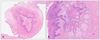
He underwent excision of a nodule on his left earlobe under the impression of neurofibroma (Fig. 1). Histological examination demonstrated a characteristic multinodular growth pattern with lobules of hyperplastic sebaceous glands forming nodules around cystically dilated follicular infundibular structures, each filled with keratin material. A prominent granular layer was seen, suggesting an infundibular type of keratinization. No hair shafts were seen in the cystic cavity. The intervening stroma showed mixed connective tissue with fibrous mesenchymal tissue, vascular channels and clusters of adipocytes. No evidence of primitive hair structures, vellus hair follicles, sweat glands or mammary ducts were seen (Fig. 2). The histopathological diagnosis of FSCH was made. After excision, no complication was observed and the wound site remained clean and healed well.
DISCUSSION
FSCH is a relatively rare form of a non-neoplastic hamartous adnexal lesion of the skin. It is relatively rare. It usually occurs on the central face and scalp, back, upper extremity, genital area and, rarely, the nipple (where there is no hair follicle)3-6. The ages of patients range from 4 to 84 years8. The name "folliculo-sebaceous cystic hamartomas" comes from the histological features: follicular cystic structure, adjacent multiple sebaceous lobules, and mesenchymal changes of the stroma.
Clinically, FSCH is usually an asymptomatic nodule or papule, usually with a rubbery to firm consistency. Although some cases of giant FSCH have been reported, the vast majority of lesions are 0.5 to 1.5 cm-sized papules or nodules9. Due to its lack of distinctive clinical features, they are often misdiagnosed as other disorders such as intradermal nevus, sebaceous hyperplasia, lipoma and neurofibroma10,11. The differential diagnosis includes STF, steatocystoma and dermoid cyst. In our case, the lesion was first diagnosed as a neurofibroma since our patient had many similar lesions on the head and neck. However, the histological findings were consistent with FSCH.
Based on histology, Kimura et al.1 suggested several criteria for diagnosis of FSCH: an infundibular cystic structure to which sebaceous lobules were attached, surrounding bundles of collagen, adipocytes, and an increased number of small venules. The differential diagnosis between FSCH and STF is challenging. Both lesions show an infundibular cyst and surrounding sebaceous nodules. However, STF has some distinctive features. Clinically, it usually presents as a depressed lesion while FSCH presents as papules or nodules. Histologically, STF exhibits hair shafts within the follicular structures while FSCH doesn't1,2. Although some authors suggest that FSCH is a late stage of STF and that histological features of FSCH are the result of secondary follicular regression of STF7, some congenital cases of FSCH cannot be explained by this theory and FSCH is generally accepted as an entity that is different from STF.
To the best of our knowledge, there is no report of FSCH in patients with neurofibroma. Different origins of FSCH and neurofibroma, and the lack of a case report suggests that the development of FSCH in a patient with neurofibroma, as in this report, is incidental.
In our case, the lack of distinctive clinical features of FSCH and the fact that the patient had NF type I led to the impression of neurofibroma. FSCH is probably more common than previously thought and it should be included in the differential diagnosis of asymptomatic skin-colored nodules or papules, especially when the lesions are located on the face and scalp.

 XML Download