

 PDF
PDF Citation
 Print
Print


INTRODUCTION
Subepidermal calcified nodule (SCN) is an uncommon form of calcinosis cutis. SCN, also called cutaneous calculi, is usually a single small and hard nodule on the face. In most instances, the surface of the nodule is verrucous; however, it may be smooth1.
SCN often occurs in damaged tissues in the absence of abnormalities in serum calcium or phosphate levels. It has been reported in a variety of local tissue injuries, including repeated trauma2. Some reports have suggested development of calcified nodules on the heel following multiple heel sticks for venesection during the neonatal period3,4. Similar cases in high risk neonates in intensive care units have been reported5-7. Though our patient had a history of heel sticks during the neonatal period, the lesion did not appear at the site where heel sticks were generally performed. Therefore, we conclude that our case is not associated with trauma.
In Korea, according to the first report on 6 cases by Son and Kim8, most lesions appeared on the eyelid and cheek8-11. There were no cases involving occurrence on the sole without trauma. We present an unusual case with unique clinical and histopathological features of childhood SCN on the sole without trauma.
CASE REPORT
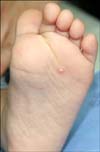
A 21-month-old boy visited our clinic because of an asymptomatic, warty papule on the left sole that had developed over the last 4 months. There was no history of preceding trauma. On physical examination, a painless, oval shaped, whitish papule measuring 5 mm on an erythematous base was observed on the left sole (Fig. 1). It resembled a verruca. The boy was otherwise in good health. Serum calcium and phosphorus levels were within normal limits.
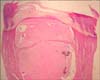
The nodule was excised by punch biopsy under local anesthesia. Haematoxylin and eosin-stained sections showed a hyperkeratotic and acanthotic epidermis overlying a cystic structure of the upper dermis and narrow pointed rete ridges in the dermis. The cystic space contained amorphous eosinophilic materials as multilobulated masses (Fig. 2). The amorphous basophilic material described in this paper appeared to be eosinophilic, indicating occurrence of degenerative and insufficient calcium on the amorphous basophilic material.
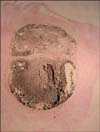
The materials were confirmed as calcium by staining with von Kossa (Fig. 3), which looks brown in color rather than black. It can be explained by two reasons: The first, the tissue sample itself showed faint staining. Early calcification, detected by von Kossa stain, could not be discovered by routine H&E staining12. The second, the high calcium area shows a black color in contrast to the brown colored calcium-lacking site. In another study of calcified lesions, 53 von Kossa-positive cases showed only 29 positive H&E (basophilic), while another 24 cases were negative on H&E13. In addition, PAS, Alcian blue (pH 2.5), and congo red stain were performed for determination of glycogen, mucopolysaccharides, and amyloid, which all showed a negative result. It suggested that the lesion contained deposits of calcium, though it appeared to show poor staining on H&E.
The lesion was completely removed by punch and there was good healing without recurrence.
DISCUSSION
In the human body, normal calcification appears only in bones and teeth. Cutaneous calcium deposits were described by Duhring as early as 1877; however, but they were first recognized by Winer14 in 1952 and named by Woods and Kellaway15 in 1963. SCN occurs more commonly in children and can also occur at birth. Incidence among male and female children is approximately equal16. Clinically, SCN is presented as a single raised, well circumscribed, whitish and hard nodule with a smooth or verrucous surface9. Multiple lesions can also be seen, but less often17. The most common location of SCN is the face18.
Pathogenesis of this disease is still unknown; however, in 1980, Tezuka19 proposed that SCN is caused by degranulation of mast cells with subsequent deposition of calcium and phosphates on the discharged mast cell contents. Histopathology of SCN consists of homogeneous basophilic masses and variable sized granules in the upper dermis with hyperkeratosis, focal parakeratosis, papillomatosis, and epidermal hyperplasia. Calcium forms granules, globules, or large masses, which often contain nuclei. A lymphohistocytic infiltrate, macrophages, foreign body granulomatous giant cells, can be seen. Calcium-containing tissue is positive for von Kossa staining18,19. Surgical excision is the treatment of choice for patients with SCN20. However, intralesional therapy (corticosteroids) has also been reported21.
In Korea, most cases of SCN occurred on the eyelid9-11 and cheek8, and, in some cases, lesions showed bilateral occurrence on thighs22, buttocks23, and fingertips24 in adulthood. Patients vary in age, from 3 to 55 years. There have been 4 cases reported as having less than a 1-year of duration of illness22,23, 1 case with 7 years8, and 2 cases with 20 years11,24. In this case, age of onset was 21 months, the youngest among domestic reports, after 4 months of spontaneous development without any trauma history. This is the age when toddling begins; therefore, we could consider the possibility that toddling itself might stimulate the onset of disease; however, there was no evidence of trauma. At the beginning, spontaneous tiny white papules without trauma were observed, and the lesion grew larger over a period of 4 months, suggesting that the lesion occurred naturally.
Our patient is asymptomatic; however, Rho et al.3 reported that this condition does not always remain asymptomatic and show spontaneous resolution. There have been several reports of calcinosis cutis classified as dystrophic calcinosis cutis, due to chronic dermal damage by multiple heel sticks in children with a medical history of admission to the neonatal intensive care unit (NICU)5-7. Development of calcinosis cutis on the sole and without trauma is rare. For this reason, we report on this case, involving asymptomatic occurrence on the sole.
 XML Download