

 PDF
PDF Citation
 Print
Print


INTRODUCTION
Proximal subungual onychomycosis (PSO) is the rarest form of onychomycosis. The pathogenic fungus invades the nail plate from the eponychium and nail matrix, and then spreads distally in PSO1. PSO initially presents as whitish patch(es) on the proximal side of the nail plate(s). At this point, we need to determine the differences between PSO and white superficial onychomycosis (WSO). Nail plates are composed of three layers, which include the dorsal, intermediate, and ventral nail plates2. Dorsal and intermediate nail plates are produced by the nail matrix, whereas the ventral nail plate is produced by the nail bed2. WSO is defined as fungal infection on the most superficial layer of the nail plate(s). Therefore, WSO shows superficial white and scaly lines or patches, which can be easily scraped from the nail plate during KOH examination. Otherwise, PSO is a fungal infection on the inner layer of the nail plate(s), because the infection starts from the eponychium and nail matrix1. Therefore, the result of KOH examination of an infected nail of PSO is almost always negative. Kaposi sarcoma is a multifocal, systemic tumor of endothelial cell origin with four clinical variants3. The variants are classic Kaposi sarcoma, endemic African Kaposi's sarcoma, Kaposi's sarcoma associated with immunosuppressive therapy, and acquired immunodeficiency syndrome (AIDS)-related Kaposi's sarcoma. Until now, no cases of Kaposi's sarcoma with onychomycosis have been reported in Korea. Herein, we report on a rare and interesting case of multiple PSO in a patient with classic Kaposi's sarcoma and suggest an easy method for KOH scraping on PSO.
CASE REPORT
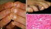
A 58-year-old man presented with whitish patches on both great toenails for four weeks prior to visiting our hospital; the patches spread rapidly to the left first to fourth finger-nails, the right third and left second to fourth toe-nails. Transverse whitish patches near the proximal nail folds were observed on finger- and toe-nails (Fig. 1A); they become broader toward the finger- and toe-tips. Prior to presentation, the patient was diagnosed with idiopathic thrombocytopenic purpura two months ago and Kaposi's sarcoma three weeks ago. The patient was treated with human immunoglobulin for five days, and then received prednisolone 40 mg bid. Upon his transfer to our hospital, we performed another biopsy on one of the multiple violaceous skin nodules in order to confirm the diagnosis of Kaposi's sarcoma (Fig. 1B). Histopathologic examination of the violaceous nodule revealed multiple slit-like spaces without endothelial lining in the dermis; these were filled with many RBCs (Fig. 1C). Testing for human herpes virus-8 (HHV-8) was not performed. The syphilis reagin test, HBs Ag, HBs Ab, and HIV Ab tests showed negative results, and results of other laboratory tests were normal, except for WBC differential count. The initial WBC count was 9.60×10⁹/L; however, percentages of segmental neutrophils, lymphocytes, monocytes, eosinophils, and basophils were 92.6%, 4%, 2.6%, 0% and 0.8%, respectively. Nail samples were obtained by scraping from superficial and deep layers of the left fourth toe-nail using a punch biopsy tool. KOH examination was performed and fungus culture was established. Fungus was found only in nail samples from the deep layer of nail plates, not in samples from the superficial nail plate.
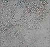
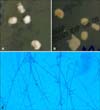
The KOH slide preparation showed long septated hyphae and numerous arthrospores (Fig. 2). The fungus culture revealed whitish downy colonies on the front side and brownish reverse pigmentation on Sabouraud's dextrose agar (Fig. 3A, B). In addition, the slide culture with lactophenol cotton-blue stain showed typical slender and clavate microconidias along the fungal hyphae (Fig. 3C). Trichophyton rubrum (T. rubrum) was isolated from fungus culture and slide culture. The internal transcribed space (ITS) regions of ribosomal DNA of the cultured fungus were sequenced, and were found to be identical to T. rubrum. The patient underwent four treatments with itraconazole (Sporanox®) pulse therapy, and was cured.
DISCUSSION
Onychomycosis denotes any infection of the nail caused by dermatophytes, nondermatophytic filamentous fungi, or yeast species4. Clinically, onychomycosis is divided into four subtypes; distal lateral subungal onychomycosis (DLSO), WSO, PSO, and total dystrophic onychomycosis (TDO). In general, PSO is known as the rarest subtype of onychomycosis. According to a study for classification of onychomycosis in Korea, the incidence of DLSO, TDO, WSO, and PSO in Korea was 76.3%, 10%, 7.9% and 5.8%, respectively5. In addition, PSO is frequently combined with immunocompromised conditions, such as HIV infection. The incidence of PSO in the immunocompetent population is 0.3%; on the contrary, that of PSO in HIV-positive individuals is 4.2~5.0%6. This might be good evidence that incidence of PSO in immunecompromised patients is higher than that in immune-competent populations. Therefore, when examining PSO patients, we should consider the possibility of immune disorders.
In general, KOH examination is an easily performed method for confirmation of onychomycosis. However, because PSO starts as white to yellow patches in the nail plate near the proximal nail fold, obtaining positive results on the intact dorsal layer of the infected nail is difficult. Nevertheless, we obtained a positive KOH examination result using the following method. Using a punch, one of the useful tools for skin punch biopsy, we made a small round incision on the infected nail plate. The site for making a round incision is on the whitish to yellowish patch(es) of the nail plate(s), and the depth of incision should be enough to remove the dorsal part of nail plate. If the depth is too shallow, a positive KOH result cannot be obtained. Therefore, the nail plate should be removed until the fragile and discolored layer of the nail plate can be seen. We then removed the most superficial layer of the nail plate and nail scraping was performed through the round hole. This method may be useful with early PSO patients who show fungal patch(es) on the nail plate(s) without an attachment to the hyponychium (free edge of the nail). In general, nail scraping for KOH examination is performed on the nail surface (in cases of WSO) or the sample is obtained from subungual hyperkeratosis (in cases of DLSO or TDO). However, in cases of PSO, nail scraping from the nail surface or subungual area can result in false negative KOH results. Therefore, new diagnostic techniques for collection of nail samples have been introduced; these include a drilling procedure and nail clipping. An article by Shemer et al7 reported interesting findings. They compared both classical technique (superficial nail scraping method) and drilling procedure. The drilling method was more superior with regard to culture sensitivity than the classical technique (drilling vs. curettage proximal, χ2=11.9, p=0.0001). English and Atkinson8 also introduced a drilling method using a drill with suction. Although the drilling method has contributed to improvement of dermatological techniques, the method has some limitations. First, the method requires use of devices, including a drill with or without suction, that are more expensive than punch tools. Second, use of the drill produces dust, which may contain fungal elements, and operators and patients can be exposed to the dust. Third, the drill makes some noise, which can cause uneasiness in patients. Finally, thickness of nail plates is varied, especially under pathologic conditions, like fungal infection. When the nail plate is very thin, it is difficult to control the depth of drilling. However, the punch method does not make any noise or dust, and the device used is very inexpensive. Furthermore, the operator can easily control the depth of incision, compared with the drilling method. Conventionally, dermatologists have used some invasive methods, such as nail extraction, in order to make a definitive diagnosis of PSO. However, nail extraction requires local anesthesia and patients need a long period of recovery. Because our newly proposed method is less invasive than the conventional method, patients do not suffer any pain, do not require any convalescent period, and the diagnosis is confirmed rapidly.
Kaposi's sarcoma is a tumor of the reticuloendothelial system, which is frequently associated with HHV-8 infection. It manifests as dark blue, violaceous plaques and nodules3 and it is common in patients with AIDS. Alteras et al9 demonstrated that fungal infections, such as tinea pedis and onychomycosis, are frequently accompanied by Kaposi's sarcoma. The microorganisms involved include T. rubrum, T. megninii, T. schoenleinii, and Epidermophyton floccosum10. Recently, several cases of altered immune status in HHV-8 associated Kaposi's sarcoma have been reported. According to one article, HIV-related and classic Kaposi's sarcoma are associated with a lack of HHV-8-specific T cells11. Although our patient did not undergo testing for HHV-8, he showed a decreased lymphocyte count. This may indicate the possibility of HHV-8 infection and decreased cellular immunity. This might be a cause of rapid spreading of PSO. However, because there is little information on the relationship between Kaposi's sarcoma and PSO, the definite cause of high incidence of PSO in Kaposi's sarcoma is still unknown.
In general, occurrence of PSO is associated with immunodeficient patients, such as AIDS patients, or organ transplant patients treated with immunosuppressive agents. On the other hand, occurrence of PSO is unusual in individuals without an immune disorder10. Dompmartin et al12 reported that more than 80% of patients with HIV infection are affected by onychomycosis. In addition, Chang and Arenas13 reported a case of PSO in a renal transplant patient who received immunosuppressive therapy. Our patient received immunoglobulin and prednisolone therapy prior to development of PSO. Several cases of PSO associated with AIDS have been reported; however, reports of PSO patients without AIDS are relatively rare. Reports of PSO with Kaposi's sarcoma without HIV infection (classic Kaposi's sarcoma) are also extremely rare. Therefore, we report on a patient with multiple PSO and classic Kaposi's sarcoma, as an interesting and unusual case.
 XML Download