

 PDF
PDF Citation
 Print
Print


INTRODUCTION
A mucocele is a benign mucous retention phenomenon that results from extravasation or retention of mucus in the surrounding tissues1. It typically presents as a translucent, bluish nodule on the lateral aspect of the lower lip. Trauma such as from biting the lip is assumed to cause most mucoceles2. On the other hand, a pyogenic granuloma is a benign, inflammatory hyperplasia of the skin and mucous membrane3. It is characterized by a localized, smooth, dome-shaped or pedunculated papule or nodule. While the pathogenesis of pyogenic granulomas remains unknown, several factors have been implicated4. Pyogenic granulomas have also been associated with trauma.
Although these lesions are both known to be associated with traumatic events, their simultaneous occurrence has only rarely been reported in the medical literature. Therefore, the purpose of this paper is to report the case of a 16-year-old female who presented to our institution with both a pyogenic granuloma and a mucocele on the lower lip.
CASE REPORT
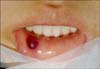
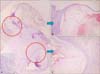
A 16-year-old female patient presented to the Department of Dermatology with a 5-month history of a 0.8×0.8 cm solitary, red-colored, polypoid nodule on the right side of the lower lip (Fig. 1). The patient had no family history of similar lesions and had no previous relevant medical history, including no history of local trauma or previous surgeries on that site. The patient complained of mild tenderness in the nodular area. The tumor was completely excised for diagnosis and treatment. On histopathologic examination, the tumor consisted of two components (Fig. 2A). The first component was a lobular structure surrounded by an acanthotic, epidermal collarette located in the upper dermis. There were multiple dilated vessels surrounded by a myxoid stroma within the lobular structure (Fig. 2B). The second component was a unilocular cyst located in the lower dermis. The tissues demonstrated infiltration of inflammatory cells and formation of granulation tissue (Fig. 2C). On the basis of clinical and histopathologic findings, the patient was diagnosed as having both a pyogenic granuloma and a mucocele. The tumor was removed completely, and there were no signs of recurrence.
DISCUSSION
A mucocele is a benign, painless, dome-shaped, soft-tissue mass that results from trauma or obstruction of the salivary gland ducts. It occurs in both males and females of any age group, but appears most frequently between the ages of 10 and 29 years1,5. The lateral aspect of the lower lip is the most common site of occurrence. However, other sites, including the upper lip and the buccal mucosa, can also be affected. Mucoceles typically present as single, recurrent, painless, well-circumscribed, bluish nodules. Most mucocles range in size from 2~10 mm in diameter. On the basis of histopathologic examination, mucoceles can be divided into two categories. The first category includes extravasation mucoceles, the more common type of mucoceles, which arise from ductal damage that causes mucus pooling in the adjacent tissue. The second category includes retention mucoceles, which result from obstruction of the excretory duct, leading to the retention of secretions and subsequent dilation of the duct. In this case, a unilocular cyst that demonstrated the typical features of an extravasation mucocele was found, as it was surrounded by inflammatory cells and granulation tissue and was located in the lower dermis. Mucoceles are best treated by excision followed by careful dissection of the affected minor salivary gland. Laser therapy and cryotherapy also can be effective treatment options6-8.
A pyogenic granuloma is a benign, inflammatory hyperplasia of the skin and mucous membrane3. The gingiva is known the most common intraoral site for the development of pyogenic granulomas. However, they also commonly affect the lips, buccal mucosa, and tongues of children and young adults9. Typically, pyogenic granulomas are solitary lesions that are smooth and dome-shaped and have the appearance of a papule or nodule. Histopathologically, they appear as lobular capillary proliferations set in a poorly cellularized, fibromyxoid stroma containing scattered inflammatory cells. In our patient, the lobular structure, surrounded by an acanthotic epidermal collarette, was observed in the upper dermis. Within the lobular structure, there were multiple dilated vessels surrounded by a myxoid stroma. Pyogenic granulomas are usually treated surgically, although cryotherapy, electrodessication, and laser therapy also have been used with varying results10.
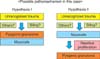
Interestingly, this case revealed the simultaneous occurrence of a pyogenic granuloma and a mucocele within a solitary polypoid nodule of the lower lip. These findings are uncommon and have rarely been reported in the medical literature. We hypothesize that two types of mechanisms might be responsible for this phenomenon (Fig. 3). The first is that both lesions developed in association with a traumatic event. Although neither lesion's etiology has been clearly identified to date, mucoceles are believed to result from trauma1,2, and pyogenic granulomas have been associated with local trauma4. On this basis, unrecognized trauma or biting could be a possible mechanism for both findings, although the patient reported no history of trauma or surgery of the lower lip. The second possible mechanism is that the pyogenic granuloma developed as a result of reactive proliferation caused by the mucocele. On the histopathologic examination, the mucocele located in the lower dermis displayed mature findings accompanying peripheral granulation tissue formation. However, the pyogenic granuloma located in the upper dermis displayed early stage lesions characterized by prominent edema without stromal fibrosis. These findings suggest that the lesions developed at different times. Because a pyogenic granuloma is an inflammatory hyperplasia that responds to various stimuli, the presence of a mucocele itself, or certain materials related to mucocele formation, might be a source of reactive proliferation leading to the development of a pyogenic granuloma9.
In summary, this case presents the possibility of a solitary lesion containing more than one source of pathology. Although we were unable to elucidate the exact pathophysiologic mechanism causing the development of both a mucocele and a pyogenic granuloma within the same lesion, either local trauma, known to be a predisposing factor for both diseases, or reactive proliferation caused by a precedent condition might explain the occurrence of this phenomenon.
 XML Download