

 PDF
PDF Citation
 Print
Print


INTRODUCTION
Acral angioosteoma cutis was first described by Googe et al.1 in 2006. They reported 11 patients with ossifying vascular lesions that were located in acral areas like the first toe, heel, finger, thumb, bottom of the foot, and palm. The lesions clinically resembled pyogenic granulomas, but their histopathological findings were not consistent with pyogenic granuloma. The authors named these novel ossifying vascular lesions "acral angioosteomas." There is one other case report about this condition2. Because acral angioosteoma cutis is rare and looks very similar to pyogenic granuloma, it is important for clinicians to have knowledge about this condition. We report a case of acral angioosteoma cutis that was completely excised with no subsequent recurrence.
CASE REPORT
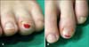
A 43-year old Korean female presented to our department for evaluation of a single 0.5×0.5 cm ulcerative, erythematous, dome-shaped subungal papule on the left fourth toe that developed after the toe bumped into a rock 18 months previously (Fig. 1). The patient's primary care physician had originally excised the lesion, but it recurred. The patient complained of mild pain but otherwise had no significant medical history or infection to the area. Laboratory values including complete blood count, calcium, phosphate, parathyroid hormone (PTH) and other electrolytes were all within reference ranges.
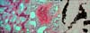
After a clinical diagnosis of cutaneous pyogenic granuloma, a deep punch biopsy and histopathologic examination were performed. The biopsy specimen showed a dilated capillary network with complete epithelial ulceration. Scattered polymorphic neutrophils and lymphocytes were also seen. Deeply eosinophilic compact materials scattered in between vascular spaces were observed. In the deeper layers, basophilic calcification foci progressively formed more definite bony trabeculae with osteocytes and blending transitions of bony components and vascular components (Figs. 2A, 2B). These bony materials exhibited positive von Kossa staining (Fig. 2C). The lobular patterns in capillary proliferation that are typical histopathological features in pyogenic granuloma were absent. The lesion was excised with electrocauterization, and a diagnosis of acral angioosteoma cutis was reached. We have followed the lesion for 12 months without observations of further aggravation or recurrence.
DISCUSSION
Cutaneous ossification can represent a heterogenous spectrum of skin disease processes. Differential diagnoses include Albright's hereditary osteodystrophy (AHO), subungal exostosis, pyogenic granuloma with metaplastic ossification, and/or osteoma cutis. AHO is characterized by short stature, a round face, and multiple skeletal abnormalities that can be seen with X-ray (curvature of the radius and shortening of the metacarpal bones). Our patient did not have the typical features of AHO. Subungal exostosis is characterized by a flesh-to-red-colored firm, immobile exophytic tumor observed most commonly in the great toe and less frequently in the other toes and fingers of young females. However, subungal exostosis has typical X-ray findings of ectopic bone formation protruding externally from the skeleton itself and has histologic features of a fibrocartilaginous cap surrounding the lesion. Pyogenic granuloma, also known as lobular capillary hemangioma, is a very common benign vascular lesion that frequently appears as a red or purple papule or polypoid mass. Extremely uncommon secondary ossification can be associated with pyogenic granuloma. We originally believed that pyogenic granuloma with ossification was the most likely diagnosis for this case. However, based on the absence of lobular capillary proliferation in the biopsy findings, pyogenic granuloma was excluded. Osteoma cutis appears very similar to cutaneous pyogenic granuloma with ectopic ossification, but the proliferation of vascular channels is not seen. Therefore, we diagnosed this case as acral angioosteoma cutis.
Acral angioosteoma cutis is a benign vascular and bony lesion occurring on the acral skin1. It is composed of well-formed capillaries, pale stroma, bland fibroblastlike cells, and multiple tiny spicules of woven bone. Lamellar bone, cartilage, or lobular arrangements of capillaries are not features of acral angiosteoma. The exact pathogenesis of acral angioosteoma cutis is unclear2. We can postulate that the pathogenesis of acral angioosteoma cutis is similar to the process of ossification in other hemagiomas. There are some reports of ossification in other kinds of hemangiomas including intramuscular hemangiomas3, skeletal muscle angiomas4, hemangiomas in the frontal sinus5, internal auditory canal6, and cavernous hemangiomas of the kidney7. However, the pathogenesis of metaplastic ossification has not been explored. There have been four documented cases of ectopic bone formation in cutaneous pyogenic granulomas to date8-11. Vascular endothelial growth factor (VEGF) and bone morphogenetic proteins (BMPs) may play a role in ectopic bone formation in pyogenic granuloma. Kim et al.10 hypothesized that trauma or infection may lead to production of VEGF, which may induce the formation of pyogenic granuloma. Hypoxia or inflammatory processes often present in pyogenic granuloma may induce expression of BMPs on endothelial cells or pericytes, which are abundant in pyogenic granuloma, and lead to osteoblastic differentiation. Our patient had a history of trauma to the area of the lesion. We believe that this trauma may have influenced the development of capillary proliferation, and added inflammation or hypoxia may have induced BMP expression. In the reported cases of pyogenic granuloma with ossification, three of four cases did not mention lobular arrangements of capillary proliferation8,9,11. Therefore, there is a possibility that some of those cases may have actually been cases of acral angioosteoma cutis.
We report an unusual case of acral angioosteoma cutis. This condition is a new entity and should be included in the differential diagnoses of cutaneous ossification diseases.
 XML Download