

 PDF
PDF ePub
ePub Citation
 Print
Print


INTRODUCTION
Acanthosis nigricans (AN) is a mucocutaneous eruption that is characterized by hyperpigmented papillomatous thickening with a velvety texture. AN can be classified into 8 variants, including the benign, obesity-associated, syndromic, malignant, acral, unilateral, medication-induced and mixed-type AN1. It usually affects the flexural area with a symmetrical distribution, but unilateral AN does not show a pattern similar to the other variants of AN2. Unilateral nevoid AN may be localized as a solitary lesion or along the line of Blaschko. Submammary involvement is quite frequent in the other variants of AN, but this has not been described in the unilateral nevoid form. We herein report the first report of submammary involvement of unilateral nevoid AN.
CASE REPORT
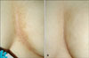
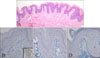
A 19-year-old female presented with asymptomatic brownish pigmented plagues on the medial side of the submammary area. Her skin lesions appeared since the age of 16 without any erythematous component and they slowly increased in size over a 3-year period. Her familial and past medical histories were unremarkable. She was neither obese nor hirsute and she had a normal menstrual cycle. On the dermatologic examination, broad linear hyperpigmented verrucous plaques were asymmetrically distributed on the intermammary to left submammary area (Fig. 1A). There were no other remarkable cutaneous or mucosal lesions. Histopathologic examination revealed hyperkeratosis, papillomatosis and moderate acanthosis in the epidermis and a slight perivascular lymphocytic infiltration in the superficial dermis (Fig. 2A). Immunohistochemical studies showed the epidermis was negative for receptors of estrogen, progesterone and androgen (Fig. 2B~D). Based on the clinical and histopathologic findings, a diagnosis of unilateral nevoid AN was made. She was treated with topical 0.025% tretinoin cream for 2 months. The skin lesions were gradually improved and they almost cleared 9 months after her first visit to our clinic (Fig. 1B).
DISCUSSION
Unilateral nevoid AN is an exceedingly rare form of AN and it was first reported as an epidermal nevus that resembled AN by Curth in 19763. Some authors have also described this dermatosis as an AN form of epidermal nevus because the lesions usually tend to be distributed unilaterally along the line of Blaschko2,3. Like our case, however, AN lesions usually have a more velvety consistency than epidermal nevus and they histopathologically show mild hyperkeratosis, papillomatosis and limited hyperplasia. Some cases of unilateral nevoid AN may be inherited in an irregular autosomal dominant manner, and the lesions manifest at birth, childhood, puberty or early adulthood. Unilateral nevoid AN has not been related to an endocrinopathy in most of the previous reports, except for two cases that were associated with Hashimoto's thyroiditis and obesity, respectively. The clinical course is unpredictable because unilateral nevoid AN can remain constant or can undergo regression1,2. The histopathologic findings are the same as the other forms of AN and the pathogenesis has been remained uncertain1-3.
Unilateral nevoid AN may simulate a hyperpigmented nevus, such as Becker's nevus and melanocytic nevus. The differential diagnosis of nevoid AN also includes confluent and reticulated papillomatosis and Dowling-Degos disease4. Yet it is not difficult to exclude these other diseases based upon the clinical and histopathologic features of unilateral nevoid AN.
Various locations of unilateral nevoid AN have been reported, including the face and scalp, chest and abdomen, and especially the periumbilical area, back and thigh2-8. However, to the best of our knowledge, no case has been described submammary involvement, which is relatively common for the other variants of AN. Because a submammary location had been observed in endocrine-related AN and the skin lesions of our case had appeared during adolescence with a clinical resemblance to Becker's nevus, we studied the expression of sex hormone receptors in the lesions to elucidate the association between sex hormones and the development of unilateral nevoid AN. It was previously reported that hyperandrogenemia played a role in the syndrome of severe insulin resistance with endocrine-related AN9 and the level of androgen receptor proteins was increased in the lesions of Becker's nevus10. In the present case, immunohistochemical studies showed the epidermis was negative for receptors of androgen, estrogen and progesterone. Our data for the sex hormones corresponds with that of most previous reports, which suggested that unilateral nevoid AN is generally not correlated with an endocrinopathy. But further studies on unilateral nevoid AN are necessary to determine the pathogenesis.
Although no guideline or consensus for the treatment of unilateral nevoid AN has been proposed due to its rare incidence, a case of ordinary AN treated with topical tretinoin was reported in 199111. Moreover, Lee et al.5 achieved clinical improvement with topical tretinoin treatment in a patient with unilateral nevoid AN. So we selected tretinoin for treating our case. The lesions were gradually flattened and the brownish pigmentation was decreased after topical treatment with tretinoin. This was probably due to shedding of hyperkeratotic squamae, as was suggested in a previous report11.
In summary, we herein report a case of unilateral nevoid AN that was distributed on the intermammary to left submammary area of a young female. Our case is the first report of submammary involvement of unilateral nevoid AN. Since sex hormone receptors were not observed on the immunohistochemical analysis, we agree with previous reports that unilateral nevoid AN is generally not associated with endocrinopathy. Furthermore, topical tretinoin is recommended as the first choice of treatment although the mechanism of action remains uncertain.
 XML Download