

 PDF
PDF ePub
ePub Citation
 Print
Print


INTRODUCTION
Eccrine syringofibroadenoma (ESFA) is a rare tumor of eccrine ductal differentiation with variable clinical findings and characteristic histological features. Five clinical subtypes have been reported: 1) multiple ESFA associated with hidrotic ectodermal dysplasia, 2) multiple ESFA without associated cutaneous features, 3) unilateral linear ESFA, 4) solitary ESFA, and 5) reactive ESFA1,2. Reactive ESFA represents an epithelial change in association with other inflammatory or neoplastic dermatoses such as chronic skin ulcers3, burn scars4, lepromatous neuropathy, venous stasis, bullous pemphigoid, erosive palmoplantar lichen planus, peristomal dermopathy, nevus sebaceous, and pre-existing malignant tumors such as sqaumous cell carcinoma. About 75 cases of ESFA have been reported in the English literature to date, and 18 of the cases (24%) were diagnosed as reactive ESFA5.
CASE REPORT
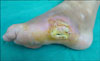
A 37-year-old woman presented with painful, multiple, coalescing, flesh-colored nodules forming a plaque on the left foot that had persisted for two years. The patient reported that the skin lesion developed several months after self-paring of a toughened area of skin on the lateral aspect of her left foot; the patient thought it was a callus. After paring, her left foot became infected, with discharge and an ulcer that was 1.5×1 cm in size. Bacterial culture of the lesion showed growth of Escherichia coli and Pseudomonas aeruginosa. The patient was admitted to the orthopedic department and was treated with antibiotics and debridement. However, the ulcer did not heal appropriately and was covered by an overlying keratotic, verrucous plaque. The plaque gradually increased in size. The patient had a history of gestational diabetes mellitus in 2006, with no other notable findings in her past. No similar lesions were reported among family members.
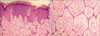
Cutaneous examination revealed multiple, coalescing, firm, flesh-colored nodules in a 'streusel bread'-like appearance, associated with areas of crusting (Fig. 1). On histopathological examination, thin anastomosing strands of uniform, small, epithelial cells arising from the epidermis to the dermis were observed (Fig. 2A). The cells were embedded in a cellular fibrous stroma and exhibited a latticed pattern characteristic of ESFA (Fig. 2B). Luminal structures were observed within the strands, and there were no cytological abnormalities. Some fungal hyphae were also observed in the corneal layer. The patient was referred to the orthopedic department and was treated with antibiotics and total excision of the cutaneous lesion.
DISCUSSION
ESFA is an uncommon tumor of eccrine glands that was first described by Mascaro6 in 1963. ESFA usually manifests as a solitary nodule on the extremities of an elderly person. Other sites of occurrence include the face, trunk and rarely the nails. Clinical findings are variable, ranging from solitary nodules to multiple papules, nodules, and plaques. Starink1 classified ESFA into four clinical subtypes: 1) multiple ESFA associated with hidrotic ectodermal dysplasia, 2) multiple ESFA without associated cutaneous features, 3) unilateral linear ESFA, 4) solitary ESFA, and French2 subsequently proposed the fifth subtype, reactive ESFA.
The diagnosis of ESFA is based on its characteristic histopathological findings. Histologically, ESFA is remarkably similar. The findings typically show proliferation of anastomosing strands and cords of monomorphous epithelial cells in a reticular pattern with eccrine duct formations embedded in a fibrovascular stroma. The histologic differential diagnosis includes fibroepithelial tumor of Pinkus, tumor of the follicular infundibulum, pseudoepitheliomatous hyperplasia, papillary eccrine adenoma, reticulated seborrheic keratosis, squamous cell carcinoma, and artifacts of histologic processing7.
It is still unclear whether the lesion is hyperplastic, hamartomatous, or neoplastic in nature. Some investigators have suggested that ESFA is a disorder that manifests as a spectrum of clinical findings, rather than being separate in nature8. The potential of progression of hyperplastic, hamartomatous, benign neoplastic, and malignant changes has been proposed, however this process has not been consecutively demonstrated in every case.
Reactive ESFA has rarely been reported in association with a chronic ulcer3 and a burn scar ulcer4. Considering our patient's history of self-paring tissue manipulation followed by infection and ulceration, reactive ESFA was the most likely diagnosis in this case. The suggested pathogenesis of reactive ESFA includes repeated eccrine duct trauma resulting in eccrine duct remodeling and repair7. Our patient demonstrates the first case of reactive ESFA in Korea.
The clinical course of ESFA is typically benign. However, malignant transformation to eccrine syringofibrocarcinoma and the association with squamous cell carcinoma has been reported in cases of ESFA9. A gradual increase in size, pain, ulcer and crust formation, and persistent lesions despite extensive treatments have been associated with malignant changes7.
The excision of the lesion has been the mainstay of treatment for solitary ESFA since malignant transformation of ESFA lesions has been reported9. Since the risk is low, however, close observation and follow-up may be an alternative to early excision, especially when complete excision is difficult due to involvement of larger areas. Since ulcers are common cutaneous findings in the dermatology department, physicians should consider ESFA and its malignant potential in the differential diagnosis of similar lesions developing from a skin ulcer.
 XML Download