

 PDF
PDF ePub
ePub Citation
 Print
Print


INTRODUCTION
Sibutramine (Reductil®) is a selective monoamine reuptake inhibitor1. It is an orally administered, centrally acting, weight-management agent devoid of amphetamine-like abuse potential. Its amine metabolites are pharmacologically active and are thought to induce the natural processes leading to enhanced satiety and thermogenesis by inhibiting serotonin and noradrenaline reuptake2. Several clinical studies assessing the effectiveness and safety of sibutramine have shown that the drug is a safe and well-tolerated agent1. Common side effects include dry mouth, constipation, nausea, and headache1. The drug has been associated with increases in blood pressure and pulse rate2. Cutaneous side effects such as urticaria, petechiae, mild skin eruption, and allergic hypersensitivity reaction have been rarely reported2. Furthermore, a case report is available on an unusual adverse reaction of erythema multiform-like bullous drug eruption caused by sibutramine3.
Drug-induced vasculitis represents approximately 10% of acute cutaneous vasculitis cases4 and is difficult to diagnose. Diagnosis and assessment of an underlying cause of a drug includes an analysis of features such as timing of drug exposure, onset, course of reaction, and nature of a recurrent eruption on rechallenge5.
Herein, we first report an interesting case of a patient with recurrent episodes of necrotizing vasculitis induced by sibutramine.
CASE REPORT
A 24-year old woman presented with hemorrhagic vesicles on her legs. The skin lesions had appeared the week before. She had been taking 10 mg sibutramine once daily for 3 months, which had been prescribed at an adequate dose and appropriate manner. She had tolerant of the drug before the development of skin lesions. She was a student, and had no relevant medical history or any viral infection history. She had no travel or trauma history.
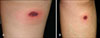
On physical examination, the patient had two overlying hemorrhagic vesicles on purple red patches on the right thigh and left calf (Fig. 1). She did not have any mucosal lesions. She was otherwise healthy, and had no history of fever, abdominal pain, or joint pain.
Kidney and liver function tests were within the normal range. C3 level was normal, and antinuclear and antineutrophil antibodies were absent. A urinalysis with microscopy showed microscopic hematuria. Kidney ultrasonography revealed a small urinary stone but otherwise normal function.
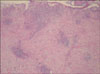
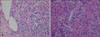
A skin biopsy from the purpuric vesicular patch revealed perivascular cellular infiltration (Fig. 2). Dense perivascular lymphohistiocytic infiltration with fibrinoid deposition in the vascular wall, red blood cell extravasation, nuclear dust, and endothelial swelling were observed revealing leukocytoclastic vasculitis (Fig. 3A). Conspicuous eosinophils were evident in the dermis (Fig. 3B). The lesions showed obvious improvement after discontinuing the sibutramine followed by the administration of 10 mg prednisolone daily for 2 weeks.
Three months later, she visited our clinic again with a recurrence of similar skin lesions on other sites of the lower extremities (Fig. 4). A history revealed that she had taken sibutramine 2 weeks before lesion recurrence. Again, the patient was told to discontinue the sibutramine, and she improved substantially within 4 days.
DISCUSSION
Vasculitis is a diverse group of segmental inflammatory disorders and blood vessel necroses. Leukocytoclastic vasculitis involves mainly small vessels in the skin and generally manifests as palpable purpura on the lower extremities6. Other features include hemorrhagic bullae, ulcers, nodules, and occasional digital necrosis6. In our case, hemorrhagic vesicles and palpable purpura were found.
Diverse histological findings are noticeable, based on the inflammation period, Red blood cell extravasation, nuclear dust, and endothelial swelling are suggestive findings of vasculitis4. At least two histological components must occur to diagnose vasculitis: a perivascular inflammatory cell infiltrate and evidence of vascular injury4. Necrosis of the vessel wall with deposition of fibrinoid material is a pathognomonic finding of vasculitis4. This patient had all vasculitis findings such as red blood cell extravasation, nuclear dust, endothelial cell swelling, perivascular cellular infiltration, and fibrinoid deposition.
When cutaneous vasculitis is demonstrated histologically, every effort should be made to define the causative agent. If visceral diseases such as renal, pulmonary, or intestinal diseases are associated, an antineutrophil antibody study should be conducted to rule out Wegener's granulomatosis, microscopic polyangitis, and Churg Strauss syndrome. If antinuclear antibody or rheumatoid factors are present and dry eye or arthritis develops, then connective tissue disease vasculitis should be considered. If urticarial lesions are found clinically, urticarial vasculitis should be diagnosed. If a high fever or other infection signs are present, then infection-related vasculitis or septic vasculitis should be considered. Paraneoplastic vasculitis should be suspected if hematological abnormalities or abnormal masses are found on imaging studies, and the lesions do not improve with corticosteroids. If there is a history of drug intake, drug-induced vasculitis should be considered.
Drug-induced vasculitis represents approximately 10% of acute cutaneous vasculitis cases5. It can be difficult to diagnose and is often a diagnosis of exclusion6. Other causes for cutaneous vasculitis, such as infection or autoimmune disease, must be excluded. Diagnosis and assessment of an underlying drug etiology consists of analyzing features such as timing of drug exposure, reaction onset, course of reaction with drug withdrawal or continuation, timing and nature of a recurrent eruption on rechallenge, and previous reports of similar reactions to the same medication6. A skin biopsy should be considered for patients with potentially severe reactions, such as those with erythroderma, blistering, skin tenderness, purpura, or pustulation6. Tissue eosinophilia may be an indicator of drug etiology in patients with cutaneous small vessel vasculitis7. The therapeutic approach can be divided into antigen removal and treatment of the cutaneous vasculitis8. Withdrawal of the precipitating medication is mandatory. Treatment for cutaneous vasculitis consists of preventing the deposition of immune complexes, and suppressing the inflammatory response8. H1 antihistamines are used to alleviate lesional symptoms and to reduce tissue deposition of circulating immune complexes. Nonsteroidal anti-inflammatory agents are combined with an H1 antihistamine, and colchicine or hydroxychloroquine sulfate can be added to or substituted for these agents8. If there is still no therapeutic response, systemic glucocorticoids or other immunosuppressants (azathioprine, methotrexate, cyclophosphamide, cyclosporine) should be considered8.
In our case, as similar skin lesions recurred after a rechallenge with sibutramine, the offending drug was sibutramine. Moreover, conspicuous eosinophilic infiltration of the tissue on histological examination suggested a drug etiology.
Sibutramine has been increasingly used since obesity has become a social issue. It is a selective monoamine reuptake inhibitor, primarily of serotonin and noradrenaline1. It is a centrally acting weight-management drug, devoid of amphetamine-like abuse potential2. Overall, selective serotonin reuptake inhibitors are safe, and adverse effects are mainly associated with gastrointestinal tract symptoms such as nausea, vomiting, or diarrhea.
Previous studies of vasculitic reactions associated with selective serotonin reuptake inhibitor therapy have been reported. Case reports of urticarial vasculitis secondary to fluoxetine9 and paroxetine10 have also been documented. Leukocytoclastic vasculitis after administering citalopram for 2 months has been reported11. A patient who developed cutaneous vasculitis while taking proxetine for 4 months has also been reported12.
Serotonin plays a role in blood clotting, evokes smooth muscle contraction, and narrows blood vessels.
Therefore, manipulating serotonin levels may result in abnormal cutaneous or visceral manifestation13. The ability of serotonin to induce and augment platelet aggregation following vascular inflammation may explain the vasculitic reaction. Further studies are needed to identify the exact mechanism of the induction of vasculitis by selective serotonin reuptake inhibitors.
No reports are available on drug-induced vasculitis associated with sibutramine. Thus, this is the first reported case of cutaneous leukocytoclastic vasculitis caused by sibutramine.
 XML Download