

 PDF
PDF ePub
ePub Citation
 Print
Print


INTRODUCTION
Pemphigus vulgaris is an autoimmune blistering disease characterized by skin and mucous membrane lesions, usually widespread and rarely localized1. If the pemphigus vulgaris lesion on the scalp persists for a long period, it may be accompanied by some inflammatory complications, such as tufted folliculitis. Moist lesions associated with pemphigus vulgaris can lead to secondary bacterial infections that cause tufting of hairs2,3.
CASE REPORT
A 51-year-old man presented to our department for an evaluation of tufted hairs accompanied by erosions and crusts on the scalp. Bullae and erosions were also observed on the trunk, both arms, both thighs, and both feet. The lesions on the scalp had been present for about 20 years, with a variable course and recurrent superinfections. The patient was prescribed medications by his prior physician for suspected seborrheic dermatitis; however, the scalp lesions did not resolve. Subsequently, multiple bullae appeared on the trunk, arms, legs, and feet 5 days before visiting our department. No additional dermatological or medical history was present.
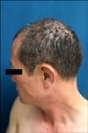
The physical examination revealed multiple irregular erosions accompanied by crusts and hair loss on the scalp; clusters of hair appeared to emerge from single follicular openings in the area with the crust (Fig. 1). In addition, flaccid bullae and vesicles were observed on the abdomen, back, both arms, both thighs, and the feet. Some bullae were ruptured and had formed erosions. No other skin or mucous membrane manifestations were present. The complete blood count showed leukocytosis; other routine blood tests were normal. Fungal cultures from the affected area of the scalp were negative, but bacterial cultures grew Staphylococcus epidermidis.
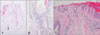
The histological evaluation of lesions on the scalp showed acantholysis with formation of intra-epidermal blisters and separation of the epidermis. Clustering of several adjacent hair follicles with perifollicular inflammatory cell infiltration was observed. A perivascular infiltrate with many lymphocytes and relatively few eosinophils was observed in the dermis (Fig. 2A, B). Another specimen obtained from the scalp showed suprabasal acantholytic cleft formation and a row of tombstone patterned cells (Fig. 2C). No spores or hyphae were noted on periodic acid Schiff staining. A direct immunofluorescence test was positive for C3 on the cell surface of keratinocytes. Additional blood samples to determine anti-dsg-1 and anti-dsg-3 antibodies were not available due to refusal by the patient. Available results led to a diagnosis of pemphigus vulgaris.
The patient was treated with local disinfectants and oral methylprednisolone. With this treatment, the lesion responded slowly and improved by the follow-up evaluation.
DISCUSSION
Although pemphigus vulgaris commonly involves the scalp, only two accounts of tufted hair folliculitis developing as a sequel of scalp involvement have been reported. Both cases involved men who had more than a year-long history of pemphigus vulgaris involving the scalp2,3. In both reported cases, Staphylococcus aureus was isolated from the lesions. In contrast, Staphylococcus epidermidis was isolated from the lesions of our patient. Some authors theorize that bacterial infection causes the tufted hair folliculitis2-5 and suggest that scalp lesions of pemphigus vulgaris are a common site of secondary bacterial infection3. An infective folliculitis leads to destruction of the upper parts of adjacent follicles leading to subsequent fibrosis that causes fibrous tissue contraction and clustering of follicular units6. Other hypotheses have been proposed for tufted hair folliculitis, such as nevoid abnormalities7 and retention of hairs in the telogen state8. These hypotheses are not defendable for pemphigus vulgaris that develops on lesions. The widespread distribution of tufted hair folliculitis provides evidence against a nevoid origin of the disorder. Numerous hair follicles in the telogen may be the result of trauma incurred by the follicles due to ongoing inflammatory processes.
A histopathological examination revealed similar findings between our case and the two reported cases. A variable number of hair shafts emerged from a single follicular infundibum, with inflammatory infiltrate observed in the upper and mid dermis. The walls of adjacent follicles were destroyed, but the follicular bulb and papillae remained intact. Similar findings were observed on histopathological examination of samples from our patient.
Tufted hair folliculitis is generally treated with systemic and topical antibiotics. Surgical excision can be effective at an early stage9. Some authors have used systemic steroids and intralesional steroids for tufted hair follicultis on pemphigus lesions2,3.
Persistent pemphigus vulgaris on the scalp may be difficult to distinguish from other diseases, including seborrheic dermatitis, tinea capitis, seborrheic pemphigoid, and some bacterial infections. Furthermore, pemphigus lesions of the scalp accompanied by tufted hairs can lead to an erroneous diagnosis and a delay in appropriate treatment. Therefore, if the lesion involves crusting, erosions, and tufted hair and persists without remission for a long time, pemphigus vulgaris must be considered as a diagnosis. This report describes an additional case of the disease, and may help increase awareness of the relationship between pemphigus vulgaris and tufted hair folliculitis.
 XML Download