

 PDF
PDF ePub
ePub Citation
 Print
Print


INTRODUCTION
Primary localized cutaneous amyloidosis is a metabolic disease of the skin that is characterized by extracellular deposition of amyloid proteins in the skin without evidence of systemic involvement. Generally, this disease is subdivided into macular, papular, and nodular type. Among these subcategories, primary localized cutaneous nodular amyloidosis (nodular amyloidosis) is the rarest type, in which dermal amyloid L deposition is presented as a single, or rarely, multiple nodules of varying size1-3. In contrast to macular or papular amyloidosis, epidermal damage is not thought to play a role in nodular amyloidosis, and the amyloid protein in nodular amyloidosis is believed to derive from immunoglobulin light chains (AL protein) produced by local plasma cells, not from degenerated keratinocytes1-6.
However, there have been a few case reports implying that an association between local trauma and deposition of AL protein may exist.
Herein, we describe a case of a 50-year-old Korean male patient with primary localized cutaneous nodular amyloidosis, which occurred following local trauma. To the best of our knowledge, this is the first report in Korea suggesting a possible triggering role of local trauma in the development of nodular amyloidosis.
CASE REPORT
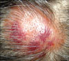
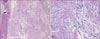
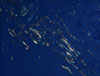
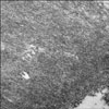
A 50-year-old Korean man with a tumefactive nodule on his frontal scalp that had increased in size over a 2 months' duration came into our clinic. He mentioned that he used to play soccer and repeatedly hit a ball mostly with the lesional frontal scalp. He recalled that he had bumped his frontal scalp against a ball rather strongly a few days before the onset of the nodule. His past medical history was unremarkable. A review of his systems found no specific symptoms. Physical examination showed a 5×5 cm sized, well-demarcated, dome-shaped, salmon-colored, waxy nodule with overlying purpuric plaques on the frontal area of the scalp (Fig. 1). The lesion was accompanied by mild tenderness. Biopsy specimens were obtained from both salmon-colored lesion and purpuric lesion. Histopathologic examination showed similar findings in both specimens. There were follicular plugging, thinning and flattening of stratum malpighii in the epidermis and deposition of acellular amorphous eosinophilicmaterials throughout the entire dermis. There were a mild to moderate perivascular and interstitial lymphocytic infiltrate admixed with plasma cells, mast cells, and a few eosinophils and a little interstitial mucin deposition in the upper and deep dermis (Fig. 2). Congo red staining with polarizing microscopic examination revealed apple-green birefringence throughout the whole dermis (Fig. 3). An electron microscopic examination revealed uniform, long, and straight filaments (Fig. 4).
Laboratory studies including complete blood cell with differential cell counts, blood chemistry, and urinalysis were within the normal limits; however, the erythrocyte sedimentation rate w as slightly elevated to 14 mm/hr (reference interval: 0~9 mm/hr). Chest radiograph showed no active lesion in the lung. Electrocardiogram showed normal sinus rhythm. Autoantibodies, including antinuclear antibody, anti-SS-A (Ro) antibody, and anti-SS-B (La) antibody, were all negative. Serum and urine protein electrophoresis analysis also produced normal results.
All these findings were consistent with a diagnosis of primary localized cutaneous nodular amyloidosis. Our patient did not want to take any specific treatment, and has remained in good general health with no evidence of progression to systemic amyloidosis and the nodule remained stable in size during a follow-up of 1 year.
DISCUSSION
Amyloid is an extracellular proteinaceous material that is amorphous, eosinophilic, homogeneous, and hyaline in microscopic appearance. Amyloidosis belongs to a metabolic disease and refers to a spectrum of conditions characterized bythedeposition of amyloid in the tissue. The deposits can be limited in the skin without evidence of systemic involvement (primary localized cutaneous amyloidosis), or ca n be systemic and involve multiple organs andtissues (primary or secondary systemic amyloidosis)2,3.
Primary localized cutaneous amyloidosis is further subdivided into macular, papular, and nodular amyloidosis. Nodular amyloidosis is the rarest type of primary localized cutaneous amyloidosis, and has somewhat distinct characteristics when compared to the other two types. A single, or rarely, multiple nodules occur preferentially on the acral areas, commonly on the face, scalp, or extremities but c an be present anywhere on the skin1-3. The nodules, which usually range from 1 to 3 cm in diameter, have a typically waxy, tumefactive appearance. Patients that are between 50 and 60 years old (mean age: 55 years) are most often affected with no sex predilection5,7.
Histopathologically, the overlying epidermis may show atrophic changes. The entire dermis and sometimes subcutis are filled with amorphous, eosinophilic, and homogeneous amyloid material. Amyloid deposits may be also found in the walls of small blood vessels and around adipocytes. A focal infiltrate of plasma cells is scattered through the deposits8,9. When stained with Congo red, amyloid deposits exhibit characteristic apple-green birefringence under polarized light. However, the above findings cannot completely exclude a diagnosis of nodular colloid degeneration. A transmission electron microscopic examination is useful to differentiate between nodular amyloidosis and nodular colloid degeneration3. Under a transmission electron microscope, amyloid filaments appear straight and long and have a uniform diameter (6 to 7 nm). This is in contrast to colloid in nodular colloid degeneration, which consists of short, wavy, and irregularly arranged filaments that are 3 to 4 nm in diameter3,10,11.
The dermal amyloid L protein is derived from immunoglobulin light chains produced by locally infiltrated plasma cells4,7,12. The precise cause of localized infiltration of plasma cells is unknown. In contrast to macular or papular amyloidosis, traumatic epidermal damage is not thought to play a role in nodular amyloidosis5. In macular or papular amyloidosis, it is strongly believed that chronic rubbing or friction of the skin leads to damage of basal keratinocytes and subsequently results in the deposits of degenerated keratinocytes as amyloid protein2. In a literature review, there are a few articles implying a possible role of local trauma in the deposition of immunoglobulin light chains. Kalajian et al.5 reported a 24-year-old woman with nodular am yloidosis on her chin, which followed local trauma from a thrown full beer can. Symonds et al.13 described a 69-year-old female patient who developed a calcifying amyloidoma (tumoral amyloidosis) on her breast following local trauma to that region. Pasternak et al.14 reported a case of soft tissue amyloidoma on the leg, where a previous traumatic event was suspected. Our case also shows a clear relationship between repeated local trauma on the frontal scalp and the onset of his nodular amyloidosis, and this raises the possibility of local trauma as a potential trigger for the development of nodular amyloidosis. More studies are required to verify the causal relationship between traumatic damage and development of nodular amyloidosis.
Although the amyloid deposits in nodular amyloidosis are composed of AL protein, the same as in primary systemic amyloidosis, by definition, nodular amyloidosis is limited to the skin without any systemic involvement and is generally a benign condition. On the basis of known association of nodular amyloidosis with systemic diseases, however, systemic evaluation and a long-term follow-up evaluation should be performed. Indeed, progression of nodular amyloidosis to systemic amyloidosis has been reported in many articles at the rate of 7% to 50%5,7,15,16. In our case, the patient had no evidence of systemic amyloidosis and laboratory findings including serum and urine protein electrophoresis, chest radiograph, and electrocardiogram were all unremarkable.
In conclusion, we describe a case of a Korean patient who developed primary localized cutaneous nodular amyloidosis after trauma. To the best of our knowledge, this is the first report in Korea suggesting a possible triggering role of traumatic damage in the development of nodular amyloidosis. However, further investigations are needed to clarify the relationship between traumatic damage and dermal amyloid L deposition.
 XML Download