This article has been
cited by other articles in ScienceCentral.
Abstract
Background
Hair dryers are commonly used and can cause hair damage such as roughness, dryness and loss of hair color. It is important to understand the best way to dry hair without causing damage.
Objective
The study assessed changes in the ultra-structure, morphology, moisture content, and color of hair after repeated shampooing and drying with a hair dryer at a range of temperatures.
Methods
A standardized drying time was used to completely dry each hair tress, and each tress was treated a total of 30 times. Air flow was set on the hair dryer. The tresses were divided into the following five test groups: (a) no treatment, (b) drying without using a hair dryer (room temperature, 20℃), (c) drying with a hair dryer for 60 seconds at a distance of 15 cm (47℃), (d) drying with a hair dryer for 30 seconds at a distance of 10 cm (61℃), (e) drying with a hair dryer for 15 seconds at a distance of 5 cm (95℃). Scanning and transmission electron microscopy (TEM) and lipid TEM were performed. Water content was analyzed by a halogen moisture analyzer and hair color was measured with a spectrophotometer.
Results
Hair surfaces tended to become more damaged as the temperature increased. No cortex damage was ever noted, suggesting that the surface of hair might play a role as a barrier to prevent cortex damage. Cell membrane complex was damaged only in the naturally dried group without hair dryer. Moisture content decreased in all treated groups compared to the untreated control group. However, the differences in moisture content among the groups were not statistically significant. Drying under the ambient and 95℃ conditions appeared to change hair color, especially into lightness, after just 10 treatments.
Conclusion
Although using a hair dryer causes more surface damage than natural drying, using a hair dryer at a distance of 15 cm with continuous motion causes less damage than drying hair naturally.
Keywords: Hair damage, Hair dryer, Heat, Natural ambient dry
INTRODUCTION
Diverse causes of extrinsic hair shaft damage have been documented, and can be roughly divided into physical causes and chemical causes
1. Chemical causes include bleaching, hair dyeing and perming. Frequent use of chemical agents is a major cause of damage to the hair shaft. When cosmetic products are used incorrectly or too frequently, they may produce changes in hair texture that correspond to morphological changes on the hair surface
2-
7. Physical causes of hair shaft damage include friction from hair accessories, washing, and towel drying. Friction is a major damage factor of the hair surface, especially in wet hair, although other factors, such as photodamage and daily grooming may also lead to hair damage. Exposure to ultraviolet radiation damages hair fibers and sunlight can lead to dryness, rough surface texture, decreased color and luster, and increased stiffness and brittleness
1,
8.
Hair dryers, which are commonly used for drying hair, also can cause hair damage. The patterns of heat damage caused by hair dryers have been investigated
9-
12. Yet, the best way to dry hair without damage remains unclear.
The purpose of this study was to observe changes in the ultra-structure, morphology, moisture content, and color of hair after repeated shampooing and drying with a hair dryer at a range of temperatures (natural ambient temperature, 47℃, 61℃, and 95℃), drying distances, and drying times.
MATERIALS AND METHODS
Hair
Chemically untreated hair was obtained from De Meo Brothers (New York, USA). The hair was washed using 1% (w/w) sodium dodecyl sulfate, and then thoroughly rinsed with tap water and dried. We selected 20 cm long hairs from the root weighing 2 g.
Hair treatments
Sodium lauryl sulfate (pH 6.0) diluted 1-in-10 was used for shampooing, and a model UN-1324B commercial hair dryer (Unix Electronics, Seoul, Korea) was used for drying. We aimed to simulate daily hair care in our experiments. In daily hair care practice, the drying temperature from hair dryer is different because of the distance between hair and hair dryer. Therefore, we After shampooing, eused different distances (5, 10, and 15 cm) between hair samples and the hair dryer. The temperature was measured 0.5 cm from the sample surface.ach hair sample was tapped gently with a towel to remove water drops. The roots were fixed to the plate and the air flow was set on the hair dryer. Each treatment was performed once a day, repeated treatments were done after 24 h. Shampooing and drying treatments were repeated 30 times for 30 days. We previously checked time to dry completely for each treatment group, so the tresses were divided into the following five test groups: (a) no treatment, (b) shampooing and drying without using a hair dryer (room temperature, 20℃), (c) shampooing and drying with a hair dryer for 60 seconds at a distance of 15 cm (47℃), (d) shampooing and drying with a hair dryer for 30 seconds at a distance of 10 cm (61℃), and (e) shampooing and drying with a hair dryer for 15 seconds at a distance of 5 cm (95℃).
Measurements
Each measurement was performed 24 h after the last treatment.
1) Scanning electron microscopy (SEM)
The prepared hair (5 cm-long from the root) was fixed onto a specimen stub and sputter-coated with gold. The hair was then inserted into a LEO 1499AP scanning electron microscope (LEO, Oberkochen, Germany) operating at an accelerating voltage of 30 kV for viewing and photography.
2) Transmission electron microscopy (TEM)
Hair was placed in propylene oxide for 15 min. After preparation with a 1 : 1 propylene oxide : Epon mixture overnight, the hair was embedded in an Epon mixture. Horizontal sections approximately 60~70 nm in thickness were cut and stained with uranyl acetate and lead citrate. The specimens were viewed with a JEM-1200EDXII transmission electron microscope (JEOL, Tokyo, Japan) operating at an accelerating voltage of 80 kV.
3) Lipid TEM
Hair was fixed in Karnovsky solution (2% glutaraldehyde+2% paraformaldehyde) and rinsed in 0.1 M sodium cacodylate and post-fixed with Lee's fixative (0.5% RuO4 : 2% OsO4 : 0.2 M cacodylate buffer = 1 : 1 : 1) at room temperature for 90 min. This procedure was designed to minimize hair injury and to better view the lipid layer of the hair. Then, each section was dehydrated in alcohol solutions substituted with propylene oxide, and embedded in the Epon mixture. The embedded section was double stained with uranyl acetate and lead citrate. Sections were examined as described for TEM.
4) Hair water content
Water content was analyzed by a HG53 halogen moisture analyzer (Mettler Toledo, Zürich, Switzerland). Individual tresses were cut to 1 cm in size and individually preserved in 82% relative humidity desiccators for 7 days before analyzing moisture content. A fragment of hair (300 mg, 1 cm in length) was placed on the saucer of the balance, and the change in weight during heating was recorded every 30 sec. The hair sample was heated for the first 40 min at 65℃, which is assumed to be the temperature of most hair dryers, and for the next 30 min at 180℃ to evaporate all water. As shown in
Fig. 1, the first converging point (A) was observed between 30 and 40 min after heating started, and the second converging point (B) was observed between 60 and 70 min after heating started. Based on the difference in weight between A and B, the second transpiring moisture content (variation of water content, %) was calculated according to the following equation: (A/A-B)×100, where A is the water content of sample after the elapsed time and B is the water content of virgin hair in each condition.
5) Color change
Hair color change was measured with a model CM-3550 spectrophotometer (Konica Minolta, Tokyo, Japan) scanning a spectral range from 360 to 740 nm in 20 nm steps. The equipment rendered CIELab values of L* (lightness), a* (red/green color axis), and b* (yellow/blue color axis). From these values, and following ASTM D 22440-85, the calculated color difference parameters were △L* (lightness difference: lighter if positive, darker if negative), △a* (red/green difference: redder if positive, greener if negative), △b* (yellow/blue difference: yellowish if positive, bluer if negative), and △E* (total color difference, △E*ab=[(△L*)2+(△a*)2+(△b*)2]½). The hair samples were measured in sets of 10.
RESULTS
Hair surface damage
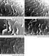
Hair surface damage was examined by SEM after repeated shampooing and drying. Lifting or cracks were not evident in the untreated and naturally dried groups (
Fig. 2A and B). In the 47℃-treated group, multiple longitudinal cracks were observed in the cuticle (
Fig. 2C). More obvious lifting and cracks of the cuticle were noted in the 61℃-treated group (
Fig. 2D). The most severe damage of the cuticle was observed in the 95℃-treated group, with many cracks, holes, and hazy cuticle borders being evident (
Fig. 2E).
Hair cuticle and cortex
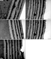
Damage to the cuticle and cortex of the hair was investigated by TEM after repeated shampooing and drying. Compared with the untreated group (
Fig. 3A), no noticeable changes were observed in naturally dried hair and the low-temperature dried hair (
Fig. 3B~D). However, punched-out cuticles were seen in the 95℃-treated group (
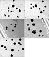
Fig. 3E). In terms of cortex damage, there were no signs of damage in any group (
Fig. 4). All cortex compartments, including melanin granules and cortical cells, were well preserved in all treated groups compared with the untreated group.
Cell membrane complex (CMC)
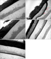
Damage to the CMC was examined by lipid TEM after the repeated shampooing and hair drying process. Only the naturally dried group exhibited the bulging that is the sign of a damaged CMC (
Fig. 5B). The CMC was well preserved with no signs of damage in control and all of the hair dryer groups (
Fig. 5A, C~E).
Moisture content analysis
Treated tresses were conditioned in a constant 82% relative humidity desiccator for 7 days and moisture content was analyzed by a halogen moisture analyzer. The changes in moisture content are summarized in
Fig. 6. Treated groups, both with and without mechanical hair drying, displayed decreased moisture content compared to the untreated group (4.6%). The moisture content was a little lower at 47℃ and 61℃ compared to 95℃ and natural drying. However, the differences between the treatment groups (group b~e) were not statistically significant. Also, differences between the control group and all treatment groups were not significant.
Color changes
Table 1 shows the changes in color under all conditions. Drying under the ambient and 95℃ conditions appeared to change hair color, especially into lightness, after just 10 treatments. In all treated groups, the hair was brighter than its original condition after 30 repeated cycles.
DISCUSSION
The human hair shaft consists of the cortex with a central axial medulla and an external cuticular layer
13. Hair damage due to heat can be found on the surface, cuticle layers, and possibly the CMC. In previous studies, common dailygrooming procedures caused more damage to the endocuticle and CMC than to other hair components
4,
9.
Repeated cycles of wetting and blow-drying can cause multiple cracks on hair cuticles
9. Other studies reported damagecaused by curling irons
10,
14. Hair shampoo surfactants and daily hair drying (including heat drying) causes damage to the ultrastructure of the hair, as well as color changes
15.
In this study, we evaluated changes to the ultra-structure, morphology, moisture content, and color of hair after repeated shampooing and drying at various temperatures (natural ambient temperature, 47℃, 61℃, and 95℃). We tried to simulate daily hair care practice, so we could suggest a pro per method to dry hair. Although the temperature of hair dryer is fixed, the temperature rises as the distance between hair and hair dryer decreases.
Hair drying without a hair dryer produced a relatively well protected hair surface, while hair that was dried using a hair dryer showed more damage of hair surfaces. In addition, the hair surfaces showed an overall tendency to become more damaged as the temperature increased, with the most severe surface damage produced after drying with the highest temperature (95℃). No cortex damage was noted in any group, suggesting that the surface of the hair might play a role as a barrier to prevent cortex damage. The cortex might be more damaged with increased repetition of the process, when the barrier of hair surface is broken. The CMC was damaged only in the naturally dried group. This result was quite unexpected, because increased temperatures generally led to more hair damage. It took over 2 h to dry the hair tress completely under ambient conditions. The hair shaft swells when in contact with water, as does the delta-layer of the CMC. The delta-layer is the sole route through which water diffuses into hair
16, and so we speculate that the CMC could be damaged when it is in contact with water for prolonged periods. Longer contact with water might be more harmful to the CMC compared to temperature of hair drying. Moisture content decreased in all treated groups (with and without the hair dryer) compared to the untreated control group. However, the differences between the groups were not statistically significant. The methods used to dry wet hair might not affect moisture content. With regard to color, the hair became lighter after repeated shampooing and drying. Drying under ambient temperatures and at 95℃ resulted in earlier changes in hair color (after just 10 treatments). The reason why the hair color is brighter after repeated shampooing and drying is unknown. On TEM examination, no decrease of melanin granules was found. However, after repeated shampooing and drying, definite damage to hair cuticle was evident on SEM examination. Therefore, we assume that the hair color change might be because of the damage to hair. Further study is needed to explain the reasons for hair color changes.
Natural drying, exposure to ambient temperature after gently remove dripping water drops with towel, is usually considered to be safer than using a hair dryer. However, damage to the CMC was noted only in the naturally dried group and earlier changes in hair color were seen in this group and the 95℃ group. This effect of natural drying has not been studied or described before. It is conceivable that a long lasting wet stage is as harmful as a high drying temperature (and may be even more dangerous to the CMC). Further evaluation about contact time with water or wet environment and hair damage is needed.
Although using a hair dryer caused more surface damage than natural drying, the results of this study suggest that using a hair dryer at a distance of 15 cm with continuous motion causes less damage than drying hair naturally.


 PDF
PDF ePub
ePub Citation
 Print
Print


 XML Download