

 PDF
PDF ePub
ePub Citation
 Print
Print


INTRODUCTION
Since elastic fiber structure and tissue distribution are key elements for understanding physical properties of skin tissues, they are important in the development of methods to study the morphology and spatial organization of elastic fibers1,2. Elastic fibers are difficult to demonstrate in routine hematoxylin-eosin (H&E) stained specimens. But under a fluorescence microscope, routinely processed and stained elastic fibers show fluorescence without any special handling3. This may be due to the binding of eosin dye to the elastic fibers. Although eosin is usually not regarded as a fluorochrome, high-fluorescence emission has been described for this dye2,4. The aim of the present study was to analyze skin elastic fibers by characterizing the eosin fluorescence detected by fluorescence microscopy and to investigate the characteristics of eosin fluorescence in degenerative elastic fibers.
MATERIALS AND METHODS
Detection of fluorescence of elastic fibers stained with H&E (eosin fluorescence microscopy)
In order to investigate the origin of fluorescence from elastic fibers stained with H&E and the detailed roles of each staining agent used in H&E staining, we stained separately normal skin sections in four different ways: unstained, hematoxylin only, eosin only, and H&E. We used a microscope which enables us to use both bright field and fluorescence microscopy. A BX50 (Olympus, Tokyo, Japan) microscope operating through a reflected light fluorescence attachment (BX-FLA) was used. Fluorescence-stained sections were examined using the following excitation/emission filter combinations (filter set for FITC): 450~480 nm exciter filter and 515 nm barrier filter and 505 nm dichroic mirror. Images were obtained by digital scientific cooled CCD camera (SPOT-RT Color model, Diagnostic Instrument, Sterling Heights, MI, USA) and SPOT imaging software.
Investigation of fluorescence findings in various human skin specimens with various histological abnormalities of the dermis
Skin specimens from thirty clinical cases with various dermal histological abnormalities, were selected for fluorescence microscopic examination of elastic fibers. Fluorescence microscopic findings were then compared with those of elastic fiber special stains to determine the relationship of the two, and to ascertain the meaning of these findings.
RESULTS
Basic features of the fluorescence of elastic fibers stained with H&E
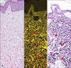
Bright yellowish fluorescence of elastic fibers, which was well differentiated with collagen fibers by their intensity and more greenish color was observed under a fluorescence microscope. In bright field microscopy of H&E-stained specimens, elastic fibers and collagen fibers were indistinguishable due to similarity in their pinkish color shades. Fluorescence findings for elastic fibers matched elastic fiber special stain (Miller's elastic stain) findings (Fig. 1).
Principle of fluorescence of elastic fibers stained in 4 different ways
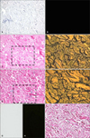
1) Fluorescence findings of unstained sections
Unstained sections of the same material were examined for verification of the contribution of autofluorescence of elastic fibers to the final image. Unstained tissue sections showed a very weak and dull green fluorescence pattern of elastic fibers. This finding was not strong enough for its adequate distinction from other structures (Fig. 2).
2) Fluorescence findings of the hematoxylin stained section
Hematoxylin stained section showed no fluorescence (Fig. 2).
3) Fluorescence findings of the eosin stained section
Eosin stained sections showed a very bright yellowish fluorescence of elastic fibers. But collagen fibers and other components (epidermis, hair) also showed excessive intensity of yellowish to greenish fluorescence enough to hamper optimal interpretation of elastic fibers. Hence, elastic fibers appeared in less definite contrast with collagen fibers (Fig. 2).
4) Fluorescence findings of the H&E stained section
In the H&E stained section, excessive eosin fluorescence of collagen fibers and other components observed in the eosin only stained section was decreased. Hence, elastic fibers appeared to have a more striking contrast with collagen fibers. Thus, hematoxylin stain quenched excessive eosin fluorescence and contributed to better contrast (Fig. 2).
Fluorescence findings of elastic fibers in the reticular and papillary dermis
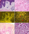
We observed some difference in findings between the fluorescence of elastic fibers in the reticular dermis and in the papillary dermis. Mature elastic fibers in the reticular dermis expressed bright yellowish fluorescence, and could be observed by fluorescence microscope. But, the thin elastic fibers (oxytalan, elaunin fibers) in the papillary dermis showed very weak fluorescence, and hence hardly ever observed by fluorescence microscopy (Fig. 3).
Investigation of fluorescence findings in various human skin specimens with suspected histological abnormalities of the dermis
1) Comparison between fluorescence microscopy of H&E-stained section and classic elastic fiber special stain
(1) Elastic fibers in sun protected skin
Fluorescence findings of elastic fibers matched well between elastic fiber special stain and fluorescence microscopy of H&E-stained section in 74% of the specimens. Usually, the matched specimens were of elastic fibers in sun-protected skin. But different elastic fiber patterns were noticed in the remaining 26% of specimens, which indicated degeneration of elastic fibers by photo aging or other causes (Table 1).
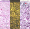
(2) Elastic fibers in photo-aged skin
Elastic fibers of sun exposed areas (face, neck and forearms) showed decreased fluorescence or no fluorescence. Hence, the elastic fibers of sun damaged skin can be distinguished from normal elastic fibers. But it is impossible to make this distinction using the classic elastic fiber special stain, because in that stain all elastic fibers are stained blue-black similarly (Fig. 4).
(3) Elastic fibers in pseudoxanthoma elasticum
The fluorescence findings of pseudoxanthoma elasticum (PXE) showed unique mixed findings of a bright yellowish fluorescence pattern of the normal elastic fibers and a mottled fluorescence pattern of the degenerative elastic fibers with calcium deposition. It is impossible to make this distinction using the classic elastic fiber special stain because in that stain all elastic fibers and calcium are stained blue-black (Fig. 5).
DISCUSSION
Elastic fibers help the skin return to its normal configuration after being stretched or deformed. The elastic fibers consist of two components: microfibrils and matrix elastin. The microfibrillar component amounts to only 15% of the elastic fiber, whereas the amorphous, electron-lucid elastin makes up 85% of the fiber5. In light microscope sections that are routinely stained, elastic fibers are inconspicuous. With special elastic tissue stains, such as orcein or resorcin-fuchisin, or in plastic-embedded sections they are found entwined among the collagen bundles6. But without any special stain, routinely processed and H&E stained elastic fibers demonstrated fluorescence under the fluorescence microscope. The fluorescence finding was related to differential binding of eosin with tissue components rather than with auto-fluorescence of the tissue components themselves2-4.
Eosin is a very common plasmal stain. It is acidic and usually employed in combination with basic dyes such as hematoxylin2. Although eosin is usually not regarded as a fluorochrome, high-fluorescence emission has been described for this dye. Eosin showed a peak emission centered at 550 nm after being excited by light of 490 nm7. And hematoxylin stain quenched excessive eosin fluorescence and contributed to better contrast8,9.
H&E staining of elastic fibers observed under fluorescence microscopy showed bright yellowish fluorescence, which would be impossible to observe under a bright field microscope. The intensity of fluorescence of elastic fibers showed a difference between the reticular dermis and the papillary dermis. Mature elastic fibers in the reticular dermis expressed bright yellowish fluorescence, and could be observed through a fluorescence microscope. But, thin elastic fibers (oxytalan, elaunin fiber) in the papillary dermis showed almost no fluorescence, and hence hardly ever observed by fluorescence microscopy. This result is consistent with the fact that the reticular dermis has more elastic fibers than the papillary dermis. It is known that the elastic system of the dermis consists superficially of thin bundles of microfibrils, which become associated with progressively larger amounts of amorphous elastin and increase in size from the deeper papillary to the reticular layer1,5. Basically, the elastic fiber system of normal human skin consists of three types of fibers: oxytalan, elaunin, and elastic fibers, which are believed to differ in their relative contents of microfibrils and elastin. According to ultrastructural analysis, oxytalan fibers contain only microfibrils, elaunin fibers contain small quantities of amorphous elastin, and elastic fibers are predominantly elastin. It is the elastin that stains with elastic tissue stains, whereas the microfibrils are the elastic resilient component of the elastic fiber1. Hence, the mature elastic fibers which are elastin-rich showed strong fluorescence. On the other hand, thin elastic fibers (oxytalan, elaunin) that had little elastin showed only very weak or almost no fluorescence.
Fluorescence images of the H&E stained sections were adequate for morphometric evaluation for elastic fibers in the dermis. And the fluorescence findings of elastic fibers matched well between elastic fiber special stain and fluorescence microscopy of the H&E-stained specimen in 74% of the specimens. But different elastic fiber patterns were found in the remaining 26% of specimens, which demonstrated degeneration of elastic fibers by photo aging or other causes. Elastic fibers of the sun-exposed areas showed decreased fluorescence or no fluorescence. Elastic fibers are acidophilic and capable of relatively selective reactions with the acid dye (eosin)10. Sun exposure induced a change in basophilic degeneration with deposition of lysozyme in the dermal elastic fiber11. This change would weaken the bonding with the acid dye (eosin), and decrease fluorescence of the elastic fibers in photo-aged skin. For this reason, elastic fibers of sundamaged skin could be distinguished from normal elastic fibers. But it has been impossible to make this kind of distinction using the classic elastic fiber special stain method.
Sometimes, analysis of skin elastic fibers by fluorescence microscopy shows better tissue morphological details than that by special stains. PXE is an inherited multi-system disorder with primary abnormalities in the elastic fibers12. Histological changes in PXE include fragmentation, clumping and calcification of elastic fibers as well as basophilic swollen and degenerated elastic fibers in the middle and deep reticular dermis13. The fluorescence findings of PXE showed unique mixed findings of a bright yellowish fluorescence pattern in the normal elastic fibers and a mottled fluorescence pattern with calcium deposition in the degenerative elastic fibers. It would be impossible to make this distinction using the classic elastic fiber special stain, because in elastic fiber special staining, all elastic fibers and calcium stain blue-black.
The present study has some limitations. We used various skin specimens to compare findings between fluorescence microscopy of H&E stained sections and classic elastic fiber special staining. Because we only used skin specimens showing histological abnormalities of the dermis, further studies on various human skin disorders regardless of dermal abnormalities are needed to elucidate the efficacy of eosin fluorescence microscopy.
In conclusion, analysis of skin elastic fibers by fluorescence microscopy is a useful and complementary method to reveal hidden elastic fibers in H&E-stained specimens without using any additional special stains and to show better tissue morphological details than that can be achieved by special stains. Application of fluorescence microscopy on routine H&E-stained specimens may unveil more useful findings and hidden information in specimens of various skin diseases in the future.

 XML Download