

 PDF
PDF ePub
ePub Citation
 Print
Print


INTRODUCTION
Nevus sebaceous (NS) is a common congenital hamartoma developing mainly on the scalp and face, as a single lesion1-4. It involves proliferative changes of the sebaceous glands, sweat glands, and the hair follicles. In childhood, it consists of a circumscribed, slightly raised plaque, in a linear arrangement or in a round shape. At puberty, the lesion becomes verrucous. A case of NS which presented as multiple lesions, occurring on the right side of the temporal scalp and on the left side of the chin, is reported in this paper.
CASE REPORT
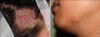
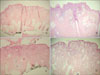
A 20-year-old man presented with a localized, slightly verrucous, hairless plaque on the right temporal scalp and a circumscribed, raised plaque on the left chin (Fig. 1). These lesions had been present since infancy. The patient wanted the plaques to be removed for cosmetic reasons. Physical examination showed that the scalp lesion was 1.5×1.2 cm in size and the face lesion was 2.0×1.5 cm. Further physical examination did not reveal any neurological, ophthalmological and cutaneous abnormalities, except for NS. There was no family history of similar skin lesions. Laboratory tests were conducted, including blood cell count, liver and renal function tests, urinalysis and venereal disease research laboratory test (VDRL), and all were within normal limits or negative. Surgical excision with a narrow safety margin was undertaken, and a primary repair with a simple suture technique was used in the scalp and the left chin. Histopathology examination revealed a large number of mature sebaceous glands and overlying papillomatous epidermal hyperplasia, without mature hair follicles (Fig. 2). At present, the patient is in the follow-up period.
DISCUSSION
The clinical and histopathological features of NS were investigated, between Jan. 1991 and Dec. 2009, in the department of Dermatology of Kyungpook National University Hospital. Among 202 cases of NS, the most common site was the scalp (66.8%), followed by the face (26.7%) and the neck (5.5%). The case reported in this study was the only patient presenting with multiple NS, i.e. on the scalp and on the contralateral side of the face. After an extensive literature review, we could not find cases of multiple NS5-8. The literature review concluded that NS commonly occurs on a single location8. However, it may be multiple and extensive, similar to verrucous epidermal nevi. Cases of multiple extensive NS, in which multiple NS never crossed the anterior sagittal midline, were reported by Correale and colleagues9. And a case of multiple NS located on both sides of the body has not been reported yet in the English literature. NS occurring on the scalp and on the ipsilateral side of the face was rarely encountered in the literature10.
In the case presented herein, the location of the second NS lesion was across the sagittal midline, i.e. on the controlateral side. Thus, one lesion occurred on the right side of the temporal scalp and the other on the left side of the chin. In addition, no multisystem disorders, such as neurological, ophthalmological and skeletal abnormalities, were associated with the linear sebaceous nevus syndrome in this case. This case presents a rare phenotype, i.e. sporadic multiple NS crossing the sagittal midline.
 XML Download