

 PDF
PDF ePub
ePub Citation
 Print
Print


INTRODUCTION
Erythema multiforme (EM) is an acute, self-limited disorder involving the skin and mucous membranes with common recurrences. EM is usually divided into two forms, EM minor and EM major. EM minor presents skin lesions without involvement of mucous membranes, while EM major is more severe, presenting both skin and mucous membrane lesions1,2.
EM affects males more often than females, with a male: female ratio ranging from 3:2 to 2:1. Although it can occur in all ages, EM usually affects adolescents and young adults, and rarely occurs during infancy and childhood. Indeed, there have been only three cases of biopsy-proven EM during the neonatal period in the literature3-7. Here, we report a case of EM in a neonate.
CASE REPORT
A 36-day-old male preterm infant (corrected age of 7 days) was admitted to our hospital with complaints that skin lesions developed on his neck and inguinal area and then spread to his entire body within a day. The patient had been born at 35 weeks and 6 days of gestation, and had a birth weight of 3,150 g. The pregnancy was uncomplicated, and there was no remarkable family history or past medical history including infections or drug intake. The first dose of hepatitis B vaccination was administered after birth, and BCG vaccination, which is generally recommended in Korea, was administered at 30 days of age.
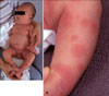
Upon admission, the infant appeared well, with only low-grade pyrexia (37.5℃) that subsided the next day. His entire body was covered with well-defined annular erythematous patches of variable size, which were typical targetoid shape. The center of the round erythematous patches was darker than the periphery (Fig. 1). However, the face, palms, soles and mucous membranes were spared. The rest of his physical examination was normal.
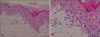
Laboratory evaluation showed neutropenia (total white blood cell [WBC] count 7.83×10³/µl; normal 5.0~19.5×10³/µl, absolute neutrophil count [ANC] 0.626×10³/µl; normal 1.0~8.5×10³/µl)8. Peripheral blood smear was conducted and revealed some anisocyotisis, poikilocytosis and lymphocytosis. However, these were not considered clinically significant. Serologic findings, including antibodies to herpes simplex virus and surface hepatitis B virus antigens, were negative. A skin biopsy specimen obtained from his right lower leg revealed a lymphohistiocytic infiltration in the upper dermis with papillary edema. Vacuolar degeneration of the basal cell layer and necrotic keratinocytes within the epidermis were also observed (Fig. 2). These findings were compatible with the diagnosis of erythema multiforme.
The patient improved rapidly upon administration of intravenous methylprednisolone at a dose of 0.5 mg/kg/day for three days, followed by a tapering dose of oral methylprednisolone over three days, and was discharged on the fifth day of admission. The skin lesions cleared without scarring within a few days; however, neutropenia persisted for a month and the ANC slowly increased. The ANC was 1.094×10³/µl one month later and 5.03×10³/µl 10 months later. At 1 year of follow up, there had been no recurrence or other systemic sequelae.
DISCUSSION
EM occurs commonly in adolescents and young adults. However, it has rarely been reported in neonates and infants3,4. Moreover, to our knowledge, there have been only three cases of biopsy-proven EM during the neonatal period, and no such cases have been reported in Korea. The first case was demonstrated by Dikland et al.5 in 1986 in a 3-week-old female. The girl had no involvement of the mucous membranes. Congenital hepatitis was diagnosed, but other microbiological and serological studies, including herpes simplex virus (HSV), showed no evidence of recent infection. The skin lesions spontaneously disappeared within two weeks without any treatment. The second case was reported by Johnston et al.6 in 2002 in a 25-day-old male. The patient had prodromal symptoms including rhinorrhea, fever, drowsiness and anorexia, which suggested an upper respiratory tract infection. He developed an unusual eruption with bullae and marked systemic symptoms. Chest X-ray revealed bilateral pulmonary infiltrates late in the course of the disease. There was no triggering factor. Symptoms began to resolve spontaneously after two weeks. The third case was described by Torrelo et al.7 in 2003 in a 2-week-old boy. The patient had no triggering factor for EM except for hepatitis B vaccination. However, hepatitis B antigens were not detected in the skin biopsy. The skin lesions cleared without scarring two weeks after the onset of the disease.
There were also several reports of neonatal EM without skin biopsy examination9-12. Suggested etiologic factors of these cases included cow's milk protein9, drugs10, hepatitis B vaccination11 and candida infection12.
In this case, the patient did not show any systemic symptoms except for transient mild fever. The short duration of fever and absence of lymphadenopathy, extremity changes and mucosal involvement excluded Kawasaki's disease. In addition, we could not find any triggering factor for EM except for hepatitis B and BCG vaccination. Considering that EM is a reactive phenomenon that occurs 5~7 days after an initial insult, BCG vaccination, which was administered five days before the onset of the disease, could have been related to development of the skin lesions. Dermatological complications after BCG vaccination are rarely seen; however, they include specific skin reactions such as lupus vulgaris, scrofuloderma, regional lymphadenitis or tuberculid and nonspecific reactions such as keloid, urticaria, granuloma annulare or erythema nodosum13,14. To date, there have been two reports suggesting an association of EM with BCG vaccination as a nonspecific reaction15,16. These reactions may be mediated by immunological hypersensitivity reaction to antigens in the vaccine. However, the exact pathogenesis remains unclear.
Interestingly, neutropenia continued for a month. The ANC was 0.626×10³/µl at admission, 1.094×10³/µl one month later, and 5.03×10³/µl ten months later. It has been reported that neonatal neutropenia occurs in 6~8% of infants, and there are many potential causes of neonatal neutropenia ranging from life threatening disease to transient benign causes of little clinical significance. Acquired or secondary neutropenia precipitated by drugs or infection is usually self-limiting. The duration of neutropenia is variable, with about 70% of the neutropenic episodes lasting less than one week but cases caused by viral infection or of unknown origin can last longer17-19. Investigations to identify the underlying infectious causes in the present case revealed no abnormality. There have been no previous studies concerning neutropenia associated with EM or BCG vaccination. We could not clarify the relationship between neutropenia and EM and thought that this finding indicated transient benign neutropenia. Although the patient had no subjective symptoms except for transient mild fever, and serologic studies revealed no evidence of infections, it might have been associated with a subclinical viral infection.
Systemic corticosteroid therapy for EM is controversial because of conflicting reports and the lack of controlled studies. Some have suggested that the use of systemic corticosteroids could decrease the severity of the disease if they were administered early during the course of the disease, while others have reported delayed recovery time and increased complications20-23.
Further studies are needed to evaluate the etiologies and treatment of neonatal EM, and dermatologists should pay attention to this rare event.
 XML Download