

 PDF
PDF ePub
ePub Citation
 Print
Print


INTRODUCTION
Keratosis pilaris (KP) is a common inherited disorder of follicular hyperkeratosis. It is characterized by small, folliculocentric keratotic papules that may have surrounding erythema1. The small papules impart a stippled appearance to the skin, resembling gooseflesh. This condition can be cosmetically very disturbing, especially in the teenagers or female patients. Topical moisturizers and keratolytics containing urea, lactic acid or salicylic acid, and topical retinoids can be used for the treatment of KP2,3. However, their effect and usefulness can be limited, due to short lasting effect and irritation. Therefore, there is a need for new and effective treatment options for the patients with KP who did not improve by the ordinary treatment. Recently, several studies in which keratosis rubra pilaris, a variant of KP, has been treated with potassium titanyl phosphate laser4 and pulsed dye laser, were reported5.
We tried to evaluate the clinical response of KP to the Q-switched 1064-nm Nd:YAG laser treatment, with regard to improvement in dyspigmentation and skin roughness. In addition, histopathologic evaluation, before and after treatment, was performed in two patients, and the adverse effects and treatment tolerability were assessed.
MATERIALS AND METHODS
Patients
This study was a pilot study to investigate the effect of the Q-switched Nd:YAG laser treatment on KP. Twelve female Korean patients with KP were enrolled. All patients were previously treated with topical agents, such as emollients, retinoids and keratolytics, but their effectiveness was limited. All of the patients were female and the mean age of the patients was 26.3 years (range: 18 to 35 years). All of the patients had bilateral symmetrical lesions on the extensor surfaces of the arms or legs and on the trunk. Before the first treatment, detailed information about the laser treatment, including risks, benefits and potential complications, was given to all enrolled patients. This study was approved by the Institutional Review Board at the Chung-Ang University Hospital. Written informed consent was obtained from all patients prior to treatment.
Procedure
The lesions were treated using a Q-switched 1064-nm Nd:YAG laser (MedLite™ C6; Hoya ConBio Inc., Fremont, CA), without local anesthesia. Ten sessions of Q-switched 1064-nm Nd:YAG laser treatment were delivered once every two weeks. All lesions were treated with the following laser settings: 4.0~5.0 J/cm2, 4-mm spot size, 10 Hz and three passes, with appropriate overlapping of the laser, until a clinical end point of mild erythema and some petechiae were observed. All subjects were also instructed to avoid sun exposure and to apply broad- spectrum sunscreen during and after the treatment.
Evaluation
1) Clinical improvement
A photograph of each patient was taken with a digital camera (EOS 40D, Canon, Japan), before each treatment session and two weeks after the last treatment. Clinical improvement was evaluated 1 month after the last treatment, using the photographic data. Two independent, experienced dermatologists evaluated the clinical improvement as seen on digital photographs taken prior to the treatment (baseline) and at 1 month after the last treatment, using a quartile grading scale (Grade 1, <25%; Grade 2, 25~50%; Grade 3, 51~75%; and Grade 4, >75% improvement) on two categories: skin texture (including the decrease in papules) and dyspigmentation (erythema and hyperpigmentation). During the study, patients were asked to report any adverse symptoms (e.g. pain, hyperpigmentation, hypopigmentation, crusting, edema, erythema, and scarring). At the end of the study, the participants documented their degree of satisfaction according to four graded-scales of very satisfied (>75%), satisfied (51~75%), slightly satisfied (25~50%), and unsatisfied (<25%).
2) Histopathologic changes
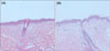
In order to evaluate the histopathologic changes, three-millimeter punch biopsies from the KP lesioned skin were obtained under local anesthesia from 2 patients with interfollicular hyperpigmentation, before the treatment and 1 month after the last treatment. Haematoxylin and Eosin (H&E) stain was used for studying the general histopathological changes in the KP lesioned skin.
RESULTS
Clinical improvement
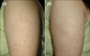
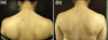
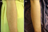
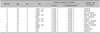
All of 12 patients completed this study, each undergoing ten sessions of Q-switched 1064-nm Nd:YAG laser treatment. After the ten sessions of treatment, the comparisons made by two independent dermatologists, between baseline and post-treatment photographs, showed that there was mild to marked improvement in all patients. In the skin texture, two patients showed >75% improvement; four showed 51~75% improvement; five showed 25~50% improvement; and only one showed <25% improvement (i.e. unsatisfied with the treatment). Regarding dyspigmentation, two patients showed >75% improvement; three showed 51~75% improvement, five showed 25~50% improvement; and only one showed <25% improvement. On the subjective self-assessments, three patients were slightly satisfied, five were satisfied, three were very satisfied, and one was unsatisfied (Table 1). Photographs showing clinical improvement after laser treatment were presented in Fig. 1, 2 and 3.
Histopathologic changes
Biopsy specimens were taken from skin on the back of patient 5 and the arm of patient 11. Before treatment, the general histopathological features of the KP specimens showed mild hyperkeratosis, perivascular mononuclear cell infiltration, and follicular plugging (Fig. 4a). After treatment, the aspect of the epidermis normalized, and follicular plugging mostly disappeared (Fig. 4b).
Adverse effects
Treatments were well tolerated without topical anesthetics. There were no significant adverse effects, such as hypopigmentation, crusting, infection, long-standing erythema, scarring, or blistering. However, some patients complained of a stinging sensation during the laser treatment, and of transient erythema after treatment. One patient complained of hyperpigmentation on the KP lesions after treatment. With time, however, hyperpigmented lesions improved without any procedures.
DISCUSSION
The KP disorder is characterized by follicular hyperkeratosis expressed as grouped folliculocentric keratotic papules, with a variable degree of perifollicular erythema1. The disorder most commonly affects the extensor aspects of the upper arms, legs, and buttocks. Patients with KP are generally asymptomatic, but occasionally may be pruritic6. Other common complaints include poor cosmetic appearance and persistent rough-textured skin, which may cause psychological distress. Treatment is usually with keratolytics, topical retinoids, as well as emollients2,3.
Control of the inflammation by topical corticosteroids also has been reported in KP4. However, these preparations, i.e. keratinolytics or topical retinoids, may aggravate the associated erythema, due to irritation, and this is limiting their value. Therefore, there is a need for new and effective treatment options for patients with KP who do not respond to conventional treatments. The pulsed tunable dye laser (PDL) treatment has been shown to be both safe and effective for the erythema of KP atrophicans7,8, which is another variant of KP. Recently, several studies of laser treatment for keratosis rubra pilaris, a variant of KP, have used either the potassium titanyl phosphate laser4 or the pulsed dye laser5. To date, to the best of the author's knowledge, there have been no published reports on the use of the Q-switched 1064-nm Nd:YAG laser therapy in the treatment of KP.
The laser device used in this study was the Q-switched 1064-nm Nd:YAG laser and the treatment parameters were short pulse durations (<10 ns), a repetition rate of 10 Hz and the use of the collimate beam mode9. Recently, this treatment method has been used for the treatment of melasma. The treatment parameters used for KP in this study were similar to those for melasma, but the energy was somewhat lower than that for dermal pigmentary lesions, such as nevus of Ota or acquired bilateral nevus of Ota-like macules.
The Q-switched 1064-nm Nd:YAG laser is a selective photothermolysis system which targets hemoglobin, water and melanin on the dermal tissue10 and might improve KP by dissolving follicular plugging and by improving erythema and pigmentation. Therefore, the Nd:YAG laser, which targets the water component, can determine follicular opening through exfoliation of the follicular plugs, subsequently improving the keratotic condition of KP. Also, we hypothesized that the 1064-nm of wave-length can penetrate deeply into the dermis, thus affecting the dermal follicular structure and resolving the follicular plugging more effectively than the conventional superficial chemical peeling agents used for KP treatment.
Some cases of KP have been associated with distinct variants of KP, i.e. erythromelanosis follicularis faciei et colli11 and KP atrophicans12. Histologically, erythromelanosis follicularis faciei et colli showed increased melanin contents of the basal layer and the upper dermis, follicular plugging, dermal vascular dilatation, congestion and perivascular inflammatory infiltration13. Thus, in erythromelanosis follicularis faciei et colli, increased melanin pigments and hemoglobin in the papillary dermal vesssels could be the chromophores targeted by the Q-switched 1064-nm Nd:YAG laser treatment. Similarly, after Q-switched Nd:YAG laser treatment, keloid and hypertrophic scars can also show clinical improvement through reduction of pigmentation and hemoglobin9. However, the precise mechanism of the therapeutic effects of the Q-switched 1064-nm Nd:YAG laser on KP is still under debate.
In this study, none of the major adverse effects of traditional laser treatments, such as permanent hypopigmentation, blistering, or scarring, were noted. Furthermore, the pain was tolerable, both during and after the procedure, even without topical anesthetics. Although this treatment modality requires multiple sessions of treatment to get clinical improvement, the improvement was maintained even at the 6-month follow-up.
In patients with KP, 1064-nm Nd:YAG laser monotherapy may not be the first line of treatment or the treatment of choice, but it could provide synergistic therapeutic effects when combined with conventional therapy. This treatment modality might be helpful, especially in patients reluctant to apply the topical agents or in those with adverse effects to the topical treatment.
In conclusion, we suggest that the Q-switched 1064-nm Nd:YAG laser might be a new promising therapeutic option for KP, without significant adverse effects.
However, this study has some limitations, such as the small number of patients and the lack of controlled group. Further well-controlled trials in a larger sample of patients are needed to fully determine the clinical effects of the Q-switched 1064-nm Nd:YAG laser on KP. In the future, additional studies will be needed to reveal the precise mechanism of action of the Q-switched 1064-nm Nd:YAG laser on KP.
 XML Download