

 PDF
PDF ePub
ePub Citation
 Print
Print


INTRODUCTION
Spitz nevus is a benign tumor that has some histological resemblance to malignant melanoma. It may be misdiagnosed as malignant melanoma and so it can be overtreated. Conversely, misdiagnosing a malignant melanoma as a Spitz nevus can be life-threatening1. In its classic clinical appearance it is colored pink-red because of the scarcity of melanin; however, tan, brown and even black pigmented Spitz nevi are also commonly found2. Spitz nevi may involve any part of the body. The head and neck area is probably the most common site of occurrence. In addition, there is a rather even distribution of lesions over the upper extremities, lower extremities and trunk3. However, an acral presentation of pigmented Spitz nevus is very rare. Herein we report on a case of acral pigmented Spitz nevus occurring on the foot and this type of lesion can clinically resemble acral lentiginous malignant melanoma.
CASE REPORT
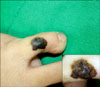
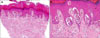
A 4-year-old male child presented to the dermatology clinic with a pigmented lesion of the left foot and the lesion had been present for 14-months. It initially started as a flat brown discoloration of the skin that gradually increased in pigmentation and size. There were no symptoms or bleeding. There was no history of excessive exposure to sunlight and no family history of cutaneous malignancy. On clinical examination, a dark brown-to black-colored, indurated plaque measuring 1×1 cm with irregular pigmentation was found on the dorsal aspect of the left fifth toe (Fig. 1). The middle of the lesion was more pigmented than the periphery. The dermoscopic examination showed multiple peripheral brownish globules and a brown to black pigmented network in the center (Fig. 1, inset). Histopathologic examination showed a symmetrical and well-circumscribed lesion that was composed of nests of epithelioid and spindle shaped melanocytes in the dermo-epidermal junction and the papillary dermis with abundant melanin and melanophages. Appropriate maturation was demonstrated as the lesion progressed in the reticular dermis. Mitosis was absent (Fig. 2). The patient was transferred to another hospital upon his parent's request even though we recommended regular follow-up or an excision.
DISCUSSION
Spitz nevus is a benign, asymptomatic, papulonodular lesion that ranges from 3 to 10 mm in diameter, and it is especially found on children and adolescents4. It usually appears as a pink or light-brown, smooth-surfaced, well-circumscribed, hairless lesion that is firm on palpation5. The Spitz nevus is classically considered to be poor in pigmentation. However, in our previous study of 80 Spitz nevi in Korean patients6, pigmentation of the melanocytes and dermal melanophages was found in 49% and 46% of the lesions, respectively, which is more common than that in previous studies, but similar to that in the Italian study by Cesinaro et al.7.
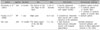
The pigmented Spitz nevus is a junctional or compound neoplasm composed of heavily pigmented, highly cohesive epithelioid and/or spindle melanocytes that are parallel and perpendicular to the skin surface, with artifactual clefts at the interface between the nests of melanocytes and the hyperplastic epidermis4. Although Spitz nevus is generally found on the skin of the face, hand, neck, trunk and lower extremities, it is rarely seen on some other parts of the body8. It has been reported that pigmented Spitz nevus is rarely localized on the acral areas of the body. Only two cases of pigmented Spitz nevus have been reported so far (Table 1). Pizzichetta et al.9 reported the first case of an acral presentation of pigmented Spitz nevus. It was in a 3-year-old boy with a single heavily pigmented, brownish black papule on the dorsum of the first finger of the left hand. They suggested two typical dermoscopic patterns (globular and starburst) of Spitz nevus might be considered different morphologic expressions corresponding to the evolutionary phases of pigmented Spitz nevus. Another case was noted on the right palmar surface of a 9-year-old girl patient10. In that previous report, dermoscopy was demonstrated to be a valuable tool for making the diagnosis of pigmented Spitz nevus, even on the glabrous skin. However, pigmented Spitz nevus was seen on the foot in our case, which has not been reported so far in the English medical literature.
Two main dermoscopic patterns of Spitz nevi have been described: (1) the starburst pattern that is characterized by prominent, black to blue diffuse pigmentation and pseudopods regularly distributed at the periphery in a radiate pattern, and (2) the globular pattern that is typified by discrete, brown to gray blue pigmentation and a peripheral rim of large brown globules that often extend throughout the entire lesion11. However, the occurrence of an atypical (multicomponent) dermoscopic pattern in Spitz nevi is well recognized, as is the occurrence of melanomas with the dermoscopic features of Spitz nevi4. This case showed an atypical dermoscopic pattern (an asymmetrically hyperpigmented lesion typified by brownish globules irregularly distributed at the periphery and a brown to black pigmented network in the center) with no histopathologic counterpart.
Histologically, Spitz nevus is more likely to contain melanin when a junctional component is present, and especially if there is no additional dermal component. The distribution of melanin is a valuable criterion for making the differential diagnosis between Spitz nevus and melanoma because when it presents, its distribution is almost always regular and superficial in Spitz nevus, whereas it is irregular and/or bottom heavy in spitzoid melanoma12. A junctional component was present in this present case. In addition, the distribution of melanin was regular and superficial.
Acral lentiginous melanoma constitutes only 2~8% of all the melanomas in Caucasians, but it is the most common form in Asians (29~46%)13. Therefore, the principal differential diagnostic consideration for a case of pigmented lesion at the acral area in Asians is acral lentiginous melanoma. Since the histologic appearance of Spitz nevus is similar to that of melanoma and the most common site of acral lentiginous melanoma is the sole, acral pigmented Spitz nevus occurring on the sole should be differentiated from acral lentiginous melanoma. As described in this case, because of the relatively large size of the lesion, it should be especially distinguished from malignant melanoma. The absence of mitosis on the histology exam helped make the differential diagnosis between the two diseases in this case. However, it would have been better to make a final definitive diagnosis of pigmented Spitz nevus after total excision rather than after a 3 mm punch biopsy in our patient to differentiate pigmented Spitz nevus from malignant melanoma. We recommended total excision, but the parents refused further follow-up.
In this report, we present a case of an acral pigmented Spitz nevus localized on the foot. To the best of our knowledge, this seems to be the first report with such localization in the medical literature. Thus, acral pigmented Spitz nevus should be considered in the differential diagnosis of pigmented lesions of the acral areas of the body, including acral lentiginous malignant melanoma.
 XML Download