

 PDF
PDF ePub
ePub Citation
 Print
Print


INTRODUCTION
Sarcoidosis is a multisystem granulomatous disorder of an unknown etiology and it is characterized by a cell-mediated immune response to a variety of exogenous antigens or autoantigens1. The lung is the most commonly affected organ, but the skin is frequently involved2. The cutaneous lesions of sarcoidosis are macules, papules and plaques that may arise as single isolated lesions or in crops. A diagnosis of sarcoidosis is made by the clinical presentation, the radiologic findings and histopathologic demonstration of noncaseating granulomas. Little is known about the association between sarcoidosis and chronic hepatitis C. According to the literature, sarcoidosis rarely occurs in hepatitis C virus (HCV) patients and it is associated with interferon (IFN) treatment1,3. Only a few cases of IFN induced sarcoidosis have been reported in the English literature3-11. IFN has an antiviral property and it has become the standard treatment for chronic HCV infection. Herein we report on a case of IFN-induced sarcoidosis that presented along the lines of venous drainage in a former intravenous drug user.
CASE REPORT
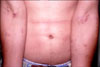
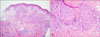
A 39-year-old male presented with multiple, violaceous papules on both antecubital areas. He'd had these lesions for the previous 2 months. The patient had been undergoing IFN-alpha monotherapy for active chronic HCV infection once in a week for the previous 6 months. He had a history of intravenous drug addiction over the previous 15 years. He complained that the skin lesions on his arms had arisen on the sites of venipuncture and IFN treatment. Several papules had also arisen on the previous injection scars, which were assumed to be injection scars because the patient was a drug addict. The papules arose along the course of the superficial veins of his arms, and several papules were dispersed on the abdomen (Fig. 1). At the same time, his review of systems was notable for an occasional cough. The chest X-ray and computed tomography scan of the patient showed that both the hilar and axillary lymph nodes were enlarged. The biopsy specimen from the forearm skin was stained with hematoxylin and eosin. Histopathological examination revealed the presence of Langhans giant cells and noncaseating, round, epithelioid granulomas in the dermis and the granulomas were circumscribed by peripheral fibrosis (Fig. 2). He was diagnosed with cutaneous and pulmonary sarcoidosis. He refused further evaluation for the systemic sarcoidosis, including assessing the level of angiotensin-converting enzyme. He received conservative treatment for the pulmonary involvement. Withdrawal of the IFN treatment and topical corticosteroid therapy for 2 weeks resulted in resolution of the cutaneous sarcoidosis.
DISCUSSION
HCV infection leads to hepatic and extrahepatic complications. Autoimmune disorders have been reported in association with HCV infection12. Essential mixed cryoglomulinemia is the most common malady associated with HCV infection, and the other reported conditions include porphyria cutanea tarda, sicca syndrome and B-cell lymphoproliferative disorders12. It has been suggested that HCV itself increases the risk of sarcoidosis7, but it appears more likely that treatment with IFN-alpha increases sarcoid granuloma formation in HCV patients. There have been three cohort studies of HCV-infected patients treated with IFN-alpha4,5,9. The prevalence of sarcoidosis in IFN-alpha treated HCV patients has been reported to range from 0.12%4 (four cases out of 3,194 patients) to 0.44%9 (3 cases out of 667 patients) in Europe. IFN-alpha activates T-helper lymphocytes and it promotes the Th1 immune response against viruses. Enhancing the Th1 immune response and macrophage activation may promote the formation of sarcoid granuloma after IFN-alpha treatment1. The proinflammatory cytokines interleukin-2 and IFN-gamma induce the expression of major histocompatibility complex II. It was recently reported that IFN-alpha polymorphism was associated with the susceptibility to developing sarcoidosis6. The clinical manifestations of IFN-induced sarcoidosis appear during antiviral therapy or more frequently after the completion of treatment4. IFN treatment induced sarcoidosis with or without ribarivin therapy. It has been suggested that the combination of IFN and ribavirin further modifies and enhances the immune response3,7,9,13. However, there have been no reported cases of sarcoidosis that occured with ribavirin-only treatment.
There has been no report of IFN-induced sarcoidosis in a Korean patient. In a review of the relevant literature via a PubMed search, the majority of cases of IFN-induced sarcoidosis in HCV patients were reported in European coutries and America4,5,7,9,13. The incidence of sarcoidosis varies widely throughout the world. The highest annual incidence of sarcoidosis has been observed in the northern European countries (5 to 40 cases per 100,000 people)14. In Japan, the annual incidence ranges from 1 to 2 cases per 100,000 people14. African Americans have about a threefold higher annual incidence as compared with that of Caucasion Americans15. Racial differences are probably influenced by environmental exposure, predisposing HLA alleles and other genetic factors1,6,11,16. HLA classes I and II are associated with sarcoidosis and IFN-induced sarcoidosis patients were positive for the predisposing HLA profile11.
Most of the cases of IFN-associated sarcoidosis reported in the literature showed a benign and uncomplicated evolution of the disease4. The clinical manifestations of IFN-induced sarcoidosis are commonly pulmonary involvement and cutaneous involvement, as compared with treatment-naive sarcoidosis in HCV patients7. Most cases of IFN induced sarcoidosis resolve spontaneously after drug discontinuation. However, approximately 11% of cases, usually those with extracutaneous involvement, had a chronic course7. IFN-induced cutaneous sarcoidosis follows a benign course and hepatitis treatment may possibly be continued with close monitoring for systemic problems such as pulmonary compromise. Some cases of cutaneous sarcoidosis resolved spontaneously despite the continuation of IFN treatment. Systemic steroids, which are the main treatment for systemic sarcoidosis, may increase the hepatitis C viral load and so the use of systemic steroids should be considered with great caution. IFN-induced sarcoidosis is a rare disease and this case is the first such report in Korea. The remarkable characteristic of our patients was the linear distribution of cutaneous sarcoidosis along the venous lines. The skin lesions on both arms had arisen at the sites of previous venipunctures, IFN treatment and scars. Although cutaneous sarcoidosis preferentially occurs within scar tissue at traumatized skin sites, there are only two cases of IFN-induced cutaneous sarcoidosis presenting along the lines of veins in the readily available English literature8,10.
Chronic hepatitis C is a worldwide disease and IFN-alpha is commonly used for eradicating the virus. Although the incidence of IFN-induced sarcoidosis in Korea is rare, we suggest that dermatologists should consider cutaneous sarcoidosis when they encounter a HCV patient who is undergoing IFN-alpha treatment and who presents with erythematous papules and nodules, and especially on previous scar sites.
 XML Download