

 PDF
PDF ePub
ePub Citation
 Print
Print


INTRODUCTION
Tumor of the follicular infundibulum (TFI) was first described in 1961, and this is a superficial epithelial plate-like proliferation of monomorphic pale-staining cells oriented in a horizontal plane with multiple thin epidermal connections1. TFI usually occurs as a solitary lesion on the head and neck of middle-aged and elderly persons. Yet TFI is rarely associated with other tumors and it presents clinically as multiple eruptive tumors. Five types of TFI have been described according to the clinical presentation: 1) solitary tumors, 2) multiple or eruptive tumors (also called infundibulomas), 3) TFI associated with typical lesions of Cowden disease, 4) TFI arising in the nevus sebaceus and 5) TFI-like epidermal changes in various tumors2. Because TFI does not have distinctive clinical features, most TFIs are diagnosed after biopsy. TFI has characteristic histopathologic features, but eccrine and sebaceous differentiation of TFI has rarely been described3,4. We report here a case of TFI with sebaceous differentiation.
CASE REPORT
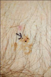
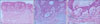
A 76-year-old man presented with a 10-year history of a 2 cm well-defined, yellowish to brownish scaly plaque on his left lower abdomen (Fig. 1). He had a history of basal cell carcinoma (BCC) on the left inguinal area, but he was otherwise healthy. Microscopic examination revealed a sharply demarcated plate-like proliferation of pale cells localized in the papillary dermis with multiple connections to the overlying epidermis (Fig. 2A). The peripheral cells showed palisading and the centrally located cells had pale-staining cytoplasm (Fig. 2B). These histopathological features were consistent with TFI. Interestingly, a cluster of mature sebaceous cells was seen in the tumor (Fig. 2C).
DISCUSSION
TFI is a rare benign adnexal tumor with characteristic histopathological features. The crucial histological findings for making the diagnosis of TFI include 1) a distinctive silhouette with horizontal proliferation, 2) characteristic neoplastic epithelial cells with small monomorphic nuclei and abundant pink cytoplasm and 3) thin columns of cells and bulkier aggregations, and all of which are interconnected5. Additionally, a dense meshwork of thin elastic fibers can be seen in the peritumoral stroma on elastic tissue staining2.
TFI is a somewhat confusing term as TFI seems to differentiate toward the isthmus rather than the infundibulum. The pale epithelial cells have been found to contain as much glycogen as the outer root sheath2. Most TFIs appear to express a cytokeratin profile comparable to that of the follicular isthmus6. Ackerman et al.5 proposed TFI usually arises from the follicular infundibulum, but it differentiates toward the isthmus of the outer sheath and toward the outer sheath in the early catagen stage.
Sebaceous differentiation is not known to be related to TFI, but a case of TFI with sebaceous differentiation has recently been reported3. The histological features of our case were similar to those of the earlier report except for the presence of colloid bodies. However, this is the first report depicting an image with the clinical features of this very rare lesion. TFI is usually skin-colored or sometimes slightly hypopigmented7. Clinically, the yellowish lesion in the current case is different from the red one in a previous report of a TFI with sebaceous differentiation. Sebaceous differentiation would explain the clinical yellowish color in this case.
Abbas and Mahalingam8 reported TFI was associated with other cutaneous lesions, including BCC, actinic keratosis, desmoplastic malignant melanoma, junctional melanocytic nevus, trichilemmoma and epidermal inclusion cyst. Our patient also had a history of BCC on the left inguinal area. This may supports the possibility of TFI representing a reactive process.
The diagnosis of TFI with sebaceous differentiation requires the exclusion of other benign epithelial tumors showing plate-like proliferation with foci of sebaceous differentiation. Superficial epithelioma with sebaceous differentiation (SESD) was foremost in the differential diagnosis. SESD is a rare benign neoplasm that is characterized by a superficial plate-like proliferation of basaloid cells with broad epidermal attachments, keratin-filled cysts and clusters of mature sebaceous cells within the tumor9,10. Generally, the presence of sebaceous differentiation favors the diagnosis of SESD rather than TFI, but it could be associated with TFI as in a previous case3. In our case, the distinction could be made by the other histopathological features. The multiple "thin" epidermal connections in our case were different from the "broad" ones of SESD2. In addition, the pale tumor cells showed the isthmus differentiation, and this favored a diagnosis of TFI with sebaceous differentiation. Additional differential diagnoses include seborrheic keratosis with sebaceous differentiation (SKSD), superficial BCC, fibroepithelioma of Pinkus, trichilemmoma and an unusual syringofibroadenoma with sebaceous differentiation2. SKSD presents as a superficial plate-like proliferation with mature sebocytes in clusters or as single cells. However, the pale tumor cells showing isthmus differentiation in our case were different from the benign squamoid cells in SKSD11. Although superficial BCC shows the formation of a fenestrated plate below the epidermis, the absence of peripheral clefting and cytologic atypia in our case was not supportive of BCC3. Fibroepithelioma of Pinkus is characterized by basaloid epithelial strands, two to three cells thick, that arise from many foci along the epidermis and the strands anastomose to compartmentalize the fibrous stroma. The superficial nature of the basaloid proliferation, the lack of atypia and the absence of a stromal fibrotic response and primitive hair germ maturation differentiated our patients' tumor from the histological characteristics of fibroepithelioma of Pinkus. As trichilemmoma has conspicuous clear cells because of their glycogen content, it can be confused with TFI. But trichilemmoma differs from TFI by the presence of distinct peripheral palisading and a hyaline eosinophilic mantle surrounding the tumor lobules. Syringofibroadenoma, unlike TFI, is characterized by the presence of a network of benign, thin epithelial cords with foci of ductal differentiation embedded in a fibrovascular stroma3.
In conclusion, we present here an unusual TFI with the clinicopathologic feature of sebaceous differentiation. Sebaceous differentiation would account for the yellowish color of the lesion.
 XML Download