Abstract
Background
Filaggrin is a key protein that facilitates the formation of skin barrier by forming a stratum corneum. Mutations in the gene encoding filaggrin (FLG) have recently been reported in patients with ichthyosis vulgaris (IV). Interestingly, there are ethnic differences between FLG mutations identified in Asians and Europeans, and few FLG mutations are overlapping between Chinese and Japanese IV patients.
Objective
The aim of this study was to investigative the genetic polymorphism of FLG in Korean IV patients.
Methods
Genomic DNA was extracted from whole venous blood specimen of Korean patients with IV and a control group, and the full sequence of FLG was determined via overlapping long-range polymerase chain reaction method.
Filaggrin is a key protein involved in the terminal differentiation of the epidermis and formation of skin barrier. The filaggrin gene (FLG) is located on human chromosome 1q21.3. It encodes the polyprotein profilaggrin, which is consisted of 10~12 tandemly repeated filaggrin subunits1,2. The aforementioned repeat number polymorphism is rarely found in other genes - a unique phenomenon in FLG. Of the 10 FLG repeat units, the eighth and tenth may possess one more repeat unit with similar base sequence, in contrast to the other repeat units, resulting 10, 11, or 12 repeat unit variants in FLG3.
Skin diseases that are attributable to the functional defects of FLG include ichthyosis vulgaris (IV; Online Mendelian Inheritance in Man (OMIM) #146700). It has long been known that IV patients have significantly reduced granular layer according to hematoxylin-eosin (H&E) staining of the patient tissue specimens. Such decrease in granular layer is due to decrease in keratohyaline granule, a key component of the granular layer. At the same time, immunohistological stain revealed reduced expression of filaggrin4.
Recently, efficient method to determine the base sequence of FLG has been developed, and some patients with IV were found to have loss-of-function mutation in FLG, indicating that FLG abnormality is involved in the pathogenesis of IV5. Based on the fact that FLG plays an important role as a skin-barrier, studies have been extended to patients with atopic dermatitis (AD), and FLG mutation was found in some cases of AD6. In addition, several studies based on European population suggested that FLG null mutations (R501X and 2282del4) predispose to other atopic disorders (asthma and allergic rhinitis). These mutations also predispose to atopic march7, early-onset AD that persists into adulthood as well as more severe asthma2,8,9.
Interestingly, distinctive differences have been found in FLG mutation between ethnic groups. Studies on FLG mutation showed that of the 27 FLG mutations reported, only two are common to Europeans and Asians10. Genetic polymorphisms (including FLG mutation) in the Koreans could be different from that in the other nations; Japanese and Chinese have more differences than similarities between them, even though they are both Asians. A recent study by Akiyama10 which examined the relationship between FLG mutation and population genetics, confirmed certain genetic variations to be unique to Europeans and Asians. FLG variations differed even between Asians. Specifically, FLG varied among Chinese11, Japanese12-14, and Taiwanese15. Only 3321delA was found to be common among Asians10.
To date, a genetic study on Korean patient was conducted that included only one subject, and the patient was demonstrated to have 3321delA, a mutation that commonly occurs in Asians16. The aim of this study was to investigate genetic polymorphisms, including FLG mutation, in Korean IV patients.
Blood samples were obtained from 7 patients with IV (<60 years of age) from independent Korean families. The diagnosis of IV was performed clinically by experienced dermatologists on the basis of clinical manifestation. Autosomal dominant pattern of inheritance was confirmed by careful history taking. The normal control group was consisted of 13 subjects who neither experienced IV nor AD. This study was approved by the Institutional Review Board of the Chung-Ang university hospital, and written informed consent was obtained from each participant prior to study entry.
Polymerase chain reaction (PCR) and sequencing analysis was performed using a protocol described in a previous study17 with some modifications. Briefly, genomic DNA from IV patients was extracted from peripheral blood samples using the G-DEX™ Genomic DNA Extraction Kit (iNtRON Biotechnology, Sungnam, South Korea). The purity of their DNA was determined using a Nanodrop ND-1000R spectrophotometer (Thermo Fisher Scientific, Wilminton, DE, USA). DNA with an optical-density ratio 260/280 of 1.8 or more was used.
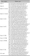
PCR amplifications were performed in 40µl reaction volumes containing 200 ng genomic DNA, 2.5 mmol/L MgCl2, 200µmol/L for each deoxynucleotide triphosphate, 10 pmol for each primer, 5xBD (Solgent Co. Ltd., Daejeon, Korea), and 2 U Taq DNA polymerase (TaKaRa Bio Inc, Otsu, Japan) using a GeneAmp PCR system 2700 (Applied Biosystems, Princeton, NJ, USA). Each genomic DNA was amplified using the primers listed in Table 1. The amplified PCR products were separated by electrophoresis in 0.8% agarose gel.
DNA sequencing was performed using Applied Biosystems 3100/3700 DNA sequencers Applied Biosystems, Princeton, NJ, USA) after conducting purification with QIAquick PCR purification kits (Qiagen, Valencia, CA, USA). Sequencing conditions were as follows: 96℃ for 1 min followed by 25 cycles of 96℃ for 10s, 50℃ for 5 sec and 60℃ for 4 min and a final extension at 10℃ for 10 min. Several sets of forward and reverse sequencing primers were used depending on the size of the PCR product fragment, as follows (Table 2).
This study was performed to determine genetic diversity (including mutation) of FLG in Korean IV patients. For the exon 1 and 2 regions, the obtained sequences were compared to the reference sequence, but no meaningful result was found. The extremely large size of exon 3, in which most of the reported mutations were found, was challenging for PCR-based sequencing method. Thus the exon 3 region was divided into 9 overlapping fragments, and PCR and sequencing analysis were carried out. Of the IV patients participated in this study, one patients had a personal and family history of AD and allergic asthma. After the full sequencing, nonsense mutation p.Y1767X (=c.5301C>G), which had never been reported in literature for IV or AD patients, was found. Later, it was revealed that his father, who had concomitant AD and asthma, carried the same mutation, in contrast to his mother who did not experience IV or AD (Fig. 1).
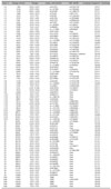
In patients from the other families, additional mutations were not found. However, 81 single nucleotide polymorphisms (SNPs) were found, more than that in the reference sequence of FLG (NG_016190.RefSeqGene). Out of 81 SNPs, 30 SNPs were new and had never been reported (Table 3). The pattern of genetic polymorphism (including repeat number polymorphism) shown by the 7 patients with IV, and by the 13 subjects in the normal control group, were compared. The chi-square test of the alleles showed no significant difference (data not shown).
After the full sequencing of FLGs in Korean IV patients and normal control, a unique diversity of the FLG gene is confirmed to exist in the Koreans. Nonsense mutation, p.Y1767X, which had never been reported, was found, and numerous SNPs that are not in the registry were observed. In some SNPs (e.g., 7th base (A>G), 310th base (C>T) of repeat 10.2, i.e. repeat after repeat 10), a homozygote of a minor allele was found in all of the subjects. This is considered to be attributable to the fact that the reference base sequence (NG_016190.1) of FLG was obtained from European populations. This justifies the necessity for the reference sequence for the Korean population.
FLG is a histidine-rich cation protein that is produced in the proteolytic degradation of profilaggrin, a large protein mass with a significant molecular weight comprised of keratohyaline granule, which exists in keratinocyte of granular layer15. In IV patients, keratohyaline granule considerably decreases or disappears, and filaggrin also decreases5,10. Filaggrin, via degradation, changes into molecules that contain urocanic acid (UCA) and pyrrolidone carboxylic acid, which block ultraviolet rays and moisturize the keratin layer15,17.
R501X and 2282del4 - mutations in the FLG genes - were first found in pediatric patients with IV who skin barrier showed abnormal functioning18. Among patients harbored either one of the two mutations, 44% of their family members have had AD, whereas among patients harbored both mutations, 76% of their family members have had AD. Among patients without any of the two mutations, no family member had AD6. A previous study showed that mutation in the FLG gene was correlated with AD of onset before the age of 2. Other studies indicate that mutations in the FLG gene that cause functional abnormality are markers of poor prognosis, and predictive that AD in an infant may manifest into adulthood19. In addition, it was also shown that the two mutations (R501X and 2282del4) that cause functional abnormalities are significantly correlated to AD (p=0.0001), asthma (p=0.006), and atopic allergy (p=0.002)12,20. Consistent with the above literature review, we could find nonsense mutation only in IV patients with concomitant personal history of AD and family history of atopic disease. This result is consistent with the previous finding for a Korean IV patient, who also experienced concomitant AD11. We found relatively lower frequency of FLG nonsense mutation in this study. One out of 7 IV patients, compared to European (ca. 50%) and other Asian populations (ca. 20%)10. We think that IV patients with personal or family history of AD should undergo investigations for FLG nonsense mutation.
The focus of previous studies was on the detection of FLG mutation in patients with IV or AD. Interestingly, however, in most of the AD patients and in a considerably large number of IV patients, loss-of-function FLG mutation did not exist. Meanwhile, reduced expression of FLG was observed in AD patients in whom such mutation was not observed21. Previous studies reported that mediators, such as sphigosylphosphorulcholine, which is involved in the inflammatory reaction of the skin, or Th2 cytokine, which is associated with pathophysiology of AD, reduced the expression of FLG22. This means that another mechanism other than FLG nonsense mutation is involved in the pathogenesis of IV or AD. Given the study finding that repeat number polymorphism can be associated with xeroderma23, we conjectured FLG length polymorphism could be an alternative explanation for most cases of IV that cannot be explalind by mutation. However, after we compared the frequency of repeat number variants between IV patients and the control group, no significant difference can not be found in the repeat number polymorphism. As for other SNPs, we could not find significant difference between IV patients and the control group. We think the so called "filaggrin-processing" (proteolytic degradation of progilaggrin to filaggrin finally leading to NMF or UCA) is another candidate to be investigated for the large portion of IV or AD patients not attributable to FLG mutations.
In summary, we established FLG full sequencing protocols applicable to the Korean population and established the Korean reference sequence for FLG. Applying these methods, we revealed previously unknown mutation in Korean IV patients and their family members. Although we could not find additional mutation or SNPs specific for Korean IV patients, we confirmed the fact that FLG nonsense mutation is likely to be found in patients with personal or family history of IV, AD, or other respiratory atopy. In the future, we hope to extend this study to Korean patients with AD, and compare the genetic diversity of FLG with other ethnic groups.
Figures and Tables
Fig. 1
Detection of filaggrin (FLG) mutation, Y1767X. (A) Direct DNA sequencing of specific FLG polymerase chain reaction products. Normal control sequence from filaggrin repeat 5 in exon3, corresponding to codons 5299-5301. Upper panels show identification of the novel Y1767X (=c.5301C>G) heterozygous mutation of the family. (B) A pedigree of IV family, showing semidominant inheritance pattern. Filled symbols refer to the IV presentation. Hetero (*): heterozygous for Y1767X, Wt: wild type for Y1767X.

References
1. Sandilands A, Terron-Kwiatkowski A, Hull PR, O'Regan GM, Clayton TH, Watson RM, et al. Comprehensive analysis of the gene encoding filaggrin uncovers prevalent and rare mutations in ichthyosis vulgaris and atopic eczema. Nat Genet. 2007. 39:650–654.

2. Barker JN, Palmer CN, Zhao Y, Liao H, Hull PR, Lee SP, et al. Null mutations in the filaggrin gene (FLG) determine major susceptibility to early-onset atopic dermatitis that persists into adulthood. J Invest Dermatol. 2007. 127:564–567.

3. Gan SQ, McBride OW, Idler WW, Markova N, Steinert PM. Organization, structure, and polymorphisms of the human profilaggrin gene. Biochemistry. 1990. 29:9432–9440.

4. McGrath JA, Uitto J. The filaggrin story: novel insights into skin-barrier function and disease. Trends Mol Med. 2008. 14:20–27.

5. Smith FJ, Irvine AD, Terron-Kwiatkowski A, Sandilands A, Campbell LE, Zhao Y, et al. Loss-of-function mutations in the gene encoding filaggrin cause ichthyosis vulgaris. Nat Genet. 2006. 38:337–342.

6. Palmer CN, Irvine AD, Terron-Kwiatkowski A, Zhao Y, Liao H, Lee SP, et al. Common loss-of-function variants of the epidermal barrier protein filaggrin are a major predisposing factor for atopic dermatitis. Nat Genet. 2006. 38:441–446.

7. Marenholz I, Nickel R, Rüschendorf F, Schulz F, Esparza-Gordillo J, Kerscher T, et al. Filaggrin loss-of-function mutations predispose to phenotypes involved in the atopic march. J Allergy Clin Immunol. 2006. 118:866–871.

8. Weidinger S, Rodríguez E, Stahl C, Wagenpfeil S, Klopp N, Illig T, et al. Filaggrin mutations strongly predispose to early-onset and extrinsic atopic dermatitis. J Invest Dermatol. 2007. 127:724–726.

9. Weidinger S, Illig T, Baurecht H, Irvine AD, Rodriguez E, Diaz-Lacava A, et al. Loss-of-function variations within the filaggrin gene predispose for atopic dermatitis with allergic sensitizations. J Allergy Clin Immunol. 2006. 118:214–219.

10. Akiyama M. FLG mutations in ichthyosis vulgaris and atopic eczema: spectrum of mutations and population genetics. Br J Dermatol. 2010. 162:472–477.

11. Ma L, Zhang L, Di ZH, Zhao LP, Lu YN, Xu J, et al. Association analysis of filaggrin gene mutations and atopic dermatitis in Northern China. Br J Dermatol. 2010. 162:225–227.

12. Nomura T, Sandilands A, Akiyama M, Liao H, Evans AT, Sakai K, et al. Unique mutations in the filaggrin gene in Japanese patients with ichthyosis vulgaris and atopic dermatitis. J Allergy Clin Immunol. 2007. 119:434–440.

13. Nomura T, Akiyama M, Sandilands A, Nemoto-Hasebe I, Sakai K, Nagasaki A, et al. Specific filaggrin mutations cause ichthyosis vulgaris and are significantly associated with atopic dermatitis in Japan. J Invest Dermatol. 2008. 128:1436–1441.

14. Nemoto-Hasebe I, Akiyama M, Nomura T, Sandilands A, McLean WH, Shimizu H. FLG mutation p.Lys4021X in the C-terminal imperfect filaggrin repeat in Japanese patients with atopic eczema. Br J Dermatol. 2009. 161:1387–1390.

15. Chen H, Ho JC, Sandilands A, Chan YC, Giam YC, Evans AT, et al. Unique and recurrent mutations in the filaggrin gene in Singaporean Chinese patients with ichthyosis vulgaris. J Invest Dermatol. 2008. 128:1669–1675.

16. Kang TW, Lee JS, Oh SW, Kim SC. Filaggrin mutation c.3321delA in a Korean patient with ichthyosis vulgaris and atopic dermatitis. Dermatology. 2009. 218:186–187.

17. O'Regan GM, Sandilands A, McLean WH, Irvine AD. Filaggrin in atopic dermatitis. J Allergy Clin Immunol. 2008. 122:689–693.
18. Sandilands A, O'Regan GM, Liao H, Zhao Y, Terron-Kwiatkowski A, Watson RM, et al. Prevalent and rare mutations in the gene encoding filaggrin cause ichthyosis vulgaris and predispose individuals to atopic dermatitis. J Invest Dermatol. 2006. 126:1770–1775.

19. Stemmler S, Parwez Q, Petrasch-Parwez E, Epplen JT, Hoffjan S. Two common loss-of-function mutations within the filaggrin gene predispose for early onset of atopic dermatitis. J Invest Dermatol. 2007. 127:722–724.

20. Weidinger S, O'Sullivan M, Illig T, Baurecht H, Depner M, Rodriguez E, et al. Filaggrin mutations, atopic eczema, hay fever, and asthma in children. J Allergy Clin Immunol. 2008. 121:1203–1209.

21. Seguchi T, Cui CY, Kusuda S, Takahashi M, Aisu K, Tezuka T. Decreased expression of filaggrin in atopic skin. Arch Dermatol Res. 1996. 288:442–446.


 PDF
PDF ePub
ePub Citation
 Print
Print


 XML Download