

 PDF
PDF ePub
ePub Citation
 Print
Print


INTRODUCTION
Lymphomatoid papulosis (LyP) is rhythmic paradoxical eruptions of erythematous papules and it has malignant histologic features and a benign clinical course1. Immunophenotyping studies of LyP lesions have shown a predominant CD4 expression. Herein we report on a case of LyP that showed a predominant CD8 expression on the immunophenotyping study.
CASE REPORT
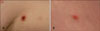
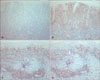
A 43-year-old man presented with a 6-month history of erythematous, asymptomatic papules on the left axilla and thigh. He had a history of hepatocellular carcinoma and this had been treated by segmentectomy of the involved portion of the liver. The family history was noncontributory. The physical examination revealed round erythematous papules with crust on the left axilla and thigh (Fig. 1). The lesions were about 1 cm in diameter. Histopathologically, there was a wedge-shaped dermal infiltrate composed of a mixture of various cell types, including lymphocytes, histiocytes, neutrophils and various atypical lymphoid cells and prominent epidermotropism (Fig. 2). The immunophenotyping revealed that the neoplastic cells were positive for CD3, CD8 and CD30, but they were negative for CD4, CD20 and CD56 (Fig. 3). The skin lesions spontaneously involuted within five months and then recurrences appeared on other sites.
DISCUSSION
LyP is classified according to the World Health Organization (WHO)-European Organization for Research and Treatment of Cancer (EORTC) as a CD30+ lymphoproliferative disorder2. Although it is not an aggressive malignant process, patients with LyP have an increased risk for developing lymphoma (10~20%) or a nonlymphoid tumour3,4. LyP is characterized by cyclic eruptions of erythematous papulonodular lesions that are usually <10 mm in size with occasional central ulceration, and this is followed by spontaneous healing and scar formation. LyP can affect patients of any age, with the peak incidence during the fifth decade1.
The most common histopathologic subtype of LyP is type A, which is characterized by atypical, large, CD30+ lymphocytes that resemble Reed-Sternberg cells of Hodgkin lymphoma, and these lymphocytes present in a wedge-shaped distribution throughout the dermis and they are admixed with various numbers of inflammatory cells. Fewer cases are classified as type B, which is characterized by small atypical lymphocytes that resemble the cerebriform cells of mycosis fungoides, and epidermotropism that is similar to that seen in the patch/plaque stage of mycosis fungoides may also be present. Type C is a rarer form of LyP. It consists of a monotonous population of large, atypical, CD30+ cells that diffusely infiltrate the dermis, with fewer associated inflammatory cells than are observed in the other types. In our case, the lesion of the axilla was consistent with type B and that of the thigh was type A. Immunohistochemically, the neoplastic cells of LyP are typically CD4+ lymphocytes that also manifest a CD30+ expression. However, rare cases of LyP that exhibited a CD8+ phenotype, as in our case, have been reported in adults and pediatric patients1,5,6.
Magro et al.5 reported on CD8+ LyP in a series of 4 patients. The lesions of these patients demonstrated the histopathologic features of CD8+ LyP and this favored a diagnosis of CD8+ LyP over one of the more characteristic CD4+ subtypes. In that report, eosinophils and neutrophils were virtually absent and striking vasculitic changes were common in this variant. There was a predominance of small uniform or monotypic lymphocytes and the presence of a granulomatous inflammation around the eccrine sweat gland coils. However, the recently reported pediatric cases showed that eosinophils and neutrophils were seen in the dermis with a polymorphic infiltration and no vasculitis or granulomas, as in our case6. Therefore, further study will be needed to establish the cardinal characteristic histopathologic findings of CD8+ LyP.
The differential diagnosis of CD8+ LyP includes primary cutaneous aggressive epidermotropic CD8+ T-cell lymphoma (CTCL) and pagetoid reticulosis2,5,7. The cornerstone for distinguishing between these disease entities is the clinicopathologic correlation. Clinically, CD8+ aggressive CTCL appears as generalized skin lesions, including erythematous-scaling patches and plaques with frequent involvement of the oral mucosa and metastatic spread to unusual sites such as the testis, lungs and central nervous system2. CD8+ aggressive CTCL show aggressive clinical behavior and a poor prognosis. Also, immunophenotyping can help make the differential diagnosis because CD8+ aggressive CTCL has commonly shown CD30-, although CD30 positivity also might occur on rare occasion5,7. On the immunophenotyping of pagetoid reticulosis, the neoplastic T cells may often be CD3+, CD4-, CD8+ and CD30 is sometimes expressed. However, pagetoid reticulosis appears as a solitary psoriasiform or hyperkeratotic patch or plaque that is usually localized on the extremities and it is slowly progressive. The typical histologic picture of pagetoid reticulosis is a hyperplastic epidermis with a marked infiltration by atypical pagetoid cells, either singly or arranged in nests. The upper dermis may show a mixed infiltrate of lymphocytes or histiocytes, but the upper dermis does not contain neoplastic T cells2. Although the long term follow-up of the previous case of CD8+ LyP was insufficient, the patients with CD8+ LyP seem to have a benign clinical course without evidence of clinical progression5. Further studies with a larger number of cases and long term follow-up are needed to estimate prognosis of CD8+ LyP and to compare it with that of CD4+ LyP.
LyP typically show a predominant CD4 expression and negativity for CD8 on immunophenotyping studies. However, a positive CD8 expression can appear in rare cases, like as in our case. CD8+ aggressive CTCL is one of representative disease entities that shows a CD8 expression and it has a poor prognosis, while CD8+ LyP has a benign clinical course. Thus, making the exact diagnosis as confirmed by the clinicopathologic correlation, as was done in our case, is important to prevent unnecessary treatment.

 XML Download