

 PDF
PDF ePub
ePub Citation
 Print
Print


INTRODUCTION
Angiokeratomas are vascular lesions defined by ectasia of the papillary dermis vessels with secondary epidermal reaction changes, such as acanthosis and/or hyperkeratosis. Clinically, they appear as one or several dark red papules, mostly with a verrucous surface1. The exact mechanism for their development is unknown; however, congenital causes, pregnancy, chilblains, trauma, and tissue asphyxia have been suggested as causal factors. Angiokeratoma circumscriptum is one of five types in the group of angiokeratomas. These lesions are usually present at birth and very few cases have been reported since its first description. Here, we report a case of angiokeratoma circumscriptum that had developed on the chest subsequently to injury.
CASE REPORT
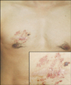
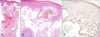
A 31-year-old man presented with asymptomatic papules on the right chest, which he had noted for twelve years prior to presentation. There was a history involving the excision of a lipoma on the right chest twelve years ago. Examination revealed several clustered, red to violaceous colored papules and plaques, measuring 2~5 mm, and distributed in a zosteriform pattern (Fig. 1). Initially, the lesions were small red macules and had been limited to the excision scar. Subsequently, the lesions became hyperkeratotic with age. The lesions were asymptomatic except for occasional episodes of minor bleeding occurring following trauma; however, these always had been easily stopped. The biopsy specimen from a representative papule demonstrated typical features of angiokeratoma (Fig. 2). The skin lesions were treated with an intense pulse light source. No recurrence has been observed during a 6 months' follow-up period.
DISCUSSION
Angiokeratomas can be classified into five types: (1) angiokeratoma of Mibelli; (2) angiokeratoma of Fordyce; (3) angiokeratoma corporis diffusum; (4) angiokeratoma circumscriptum; and (5) solitary or multiple angiokeratomas, which are similar to the Mibelli type, smaller than angiokeratoma circumscriptum, and can occur in any part of body2,3.
The histological findings are identical for all five aforementioned types of angiokeratoma and consist of dilated, thin-walled, congested capillaries mainly in the papillary dermis overlying an epidermis that exhibits acanthosis and hyperkeratosis4.
Angiokeratoma circumscriptum is the least frequent among the five types of angiokeratomas5, and its lesions are usually present at birth and may be associated with nevus flammeus, cavernous hemangioma, or hemangiectic hypertrophy. Although they are present at birth as a small erythematous macular area, there is no obvious hereditary pattern6. These lesions, as reported by Imperial and Helwig3, are not true angiomas but rather, represent telangiectasias of preexisting vessels. The basic pathological process involves dilatation of the papillary capillaries with secondary epidermal changes as a means of preventing further dilatation and rupture. These are thought to be a vascular lesions arising from local damage of papillary capillaries; however, there have been no reports of angiokeratoma circumscriptum arising from injury. In contrast, one unique case of solitary angiokeratoma developed after injury7.
Clinically, angiokeratoma circumscriptum is characterized by aggregates of hyperkeratotic erythematous papules and nodules, which may coalesce to form verrucous plaques. Lesions most commonly involve the legs in a unilateral or otherwise asymmetrical distribution; however, involvement of the trunk may also occur and in one unique case, only the neck was involved.
Differential diagnosis includes verrucous hemangioma, Cobb syndrome, lymphangioma circumscriptum, and certain tumors, including melanomas. Verrucous hemangioma is a congenital, localized vascular malformation, histologically characterized by dilated capillaries extending into the deep dermis and hypodermis.
The treatment of larger lesions in angiokeratoma usually requires laser management given that other treatment modalities may produce extremely disfiguring results8. Several laser sytems have been used to treat angiokeratomas, including argona lasers, carbon-dioxide lasers, Cooper vapor lasers, neodymium: yttrium-aluminumgarnet (Nd:YAG) lasers, pulsed dye lasers, and intense pulse light source systems.
Herein we have reported a rare case of angiokeratoma circumscriptum following the excision of a lipoma on the right chest twelve years prior. Therefore, this case indicates that injury could be involved in the development of the rare condition, angiokeratoma circumscriptum.
 XML Download