

 PDF
PDF ePub
ePub Citation
 Print
Print


INTRODUCTION
Nevus depigmentosus (ND) is a congenital, non-progressive, hypopigmented lesion that is usually stable throughout an affected individual's lifetime. The clinical features of vitiligo are similar to those of ND, but the two diseases have different treatment responses and prognoses. We report here on a rare case of vitiligo that was coexistent with ND. Both conditions were treated with narrow-band UVB.
CASE REPORT
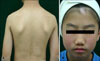
A stable hypopigmented patch was detected on the back of an 11-year-old Korean boy at birth and it had slowly enlarged as he grew older (Fig. 1A). The child incidentally discovered a hypopigmented macule with leukotrichia on his forehead 3 months before he presented at our clinic (Fig. 1B). One year earlier, he had noted pruritic scaly hypopigmented macules on his upper eyelid and cheek. The lesions disappeared after treatment with topical steroids, but they relapsed after treatment was discontinued (Fig. 1B). The boy was in good general health and all the lesions were asymptomatic. He had neither a history of trauma nor a family history of such lesions. Wood's light examination of the lesion on his back showed no enhancement, whereas Wood's light accentuated his forehead lesion. On the physical examination, he showed hypopigmented patches on his right lower back and forehead.
The results of the routine laboratory examinations, including chest posterior-anterior X-ray, the complete blood count, the liver function test, and urine analysis, were within the normal limits. The potassium hydroxide mounts prepared from both hypopigmented lesions were negative. All the procedures were performed by a single investigator under controlled ambient conditions (room temperature 22°C and relative humidity 42%). A Mexameter® (Courage- Khazaka Electronic, Koln, Germany) was used to measure the melanin index (MI) of both lesions. The MI of the back lesion was 154 and that of the forehead lesion was 83. To eliminate the confounding effect of regional skin color differences, the relative melanin index (RMI) was calculated as follows: RMI (%)=MI of lesion×100/MI of a symmetrically located normally pigmented area. The RMI of the back lesion was 66.96% and that of the forehead lesion was 30.74%.
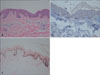
Incisional biopsy was performed on the margin of the lesion and this included some of the adjacent normal tissue. Histopathological examination of the back lesion revealed normal melanocytes in the basal layer, and this result was confirmed with S-100 protein immunoperoxidase staining (Fig. 2A, B). On the Fontana-Masson stain, the basal layer on the left side of the slide was decreased in thickness compared to that of the normal skin on the right side (Fig.2C).
Based on the clinical course (appearance, non-progressive), the lack of fluorescence under Wood's light, a higher RMI than the forehead lesion and the histopathological characteristics, the lesion on the patient's back was diagnosed as ND. The hypopigmented forehead patch, which was characterized by leukotrichia, an increase in size, accentuation under Wood's light and a lower RMI, was diagnosed as vitiligo.
After the diagnosis, treatment with narrow-band UVB was initiated once or twice a week at 200 mJ/cm2 and this was halted after 32 exposures with a total cumulative dose of 3,800 mJ/cm2. If the radiation dose administered the previous week was tolerated, then it was increased by 10% at the subsequent treatment session. If the previous treatment resulted in erythema, then no treatment was given at the next scheduled visit.
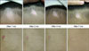
After 1, 2, 4 and 7 months, both lesions showed significant repigmentation. The vitiligo lesion on the child's forehead dramatically decreased in size and it began to show repigmentation at 2 months (Figs. 3 and 4). In contrast, the ND lesion on the child's back began to show repigmentation at 4 onths and it did not change in size
(Figs. 3 and 4).
DISCUSSION
Vitiligo is an acquired, progressive, hypopigmented disease, whereas ND is congenital and non-progressive and it is a hypopigmented lesion that is stable in size1. Segmental vitiligo, in particular, tends to show an earlier onset and it remains more stable than generalized vitiligo, and it overlaps with the clinical features of ND to a greater degree. Thus, the differential diagnosis of ND versus vitiligo must rely on the medical history, the physical examination, the Wood's light exam and histopathological evaluation2.
Several devices have recently been introduced to measure the amount of melanin pigment in the skin. Although these devices are often used for cosmetics research, they have only rarely been applied in clinical practice3,4. In this study, we used the Mexameter®, a narrow-band reflectance spectrophotometer, to measure the intensity of the erythema and melanin pigmentation. Previous studies have demonstrated that the Mexameter® is highly discriminative and sensitive enough to detect small differences in skin color and to provide satisfactory measurement reproducibility4. Park et al.2 reported that the MI, as measured using a Mexameter®, is significantly higher in ND lesions (138.17±43.87) than in vitiligo lesions (79.11±40.24). However, the MIs of vitiligo lesions overlap with those of ND, possibly due to the presence of residual melanocytes in the former, as well as other possible factors such as variations in the skin type and test site. Thus, Park et al.2 introduced the RMI, which represents the ratio of pigmentation at the lesion site relative to that of a symmetrically located normal site. As expected, the mean RMI of vitiligo lesions (50±24%) was also significantly lower than that of ND lesions (74±13%). All the ND lesions had an RMI>50%, suggesting that vitiligo can be defined as any lesion with an RMI≤50%. Similarly in our patient, the RMI of the ND lesion was 66.96% and that of the vitiligo lesion was 30.74%2.
A few therapeutic attempts have been aimed at achieving the repigmentation of ND sites by surgical methods. However, unlike vitiligo treatment, surgery has a limited role in ND treatment because the extent of repigmentation from graft foci depends on the migration of melanocytes and this is difficult to predict in these cases5. There has been only one report of repigmentation of a ND lesion following treatment with 308-nm excimer laser irradation6. Our patient was treated for ND and vitiligo with narrow-band UVB irradiation, which successfully led to the repigmentation of both lesion sites. ND is thought to be caused by functional defects in melanocytes and morphological abnormalities in the melanosomes1. A recent report suggested that UV radiation induces the up-regulation and activation of compounds that increase the phagocytosis of melanosomes, such as protease-activated receptor 27. In our patient, UV radiation also exerted a positive effect on ND, perhaps by disrupting melanosomal transfer. Prior to this report, only one other case of a patient with both vitiligo and ND had been published in the medical literature in 1996. For that patient, both lesions were treated with topical and systemic steroids. In that previous case, the vitiligo lesion showed repigmentation, but the ND lesion did not8.
Although the long-term success of narrow-band UVB for the treatment of ND has not been evaluated, this approach provided partial resolution of both the ND and vitiligo on our patient.
 XML Download