

 PDF
PDF ePub
ePub Citation
 Print
Print


INTRODUCTION
Acanthosis nigricans is prevalent in the general population and is the most common dermatological manifestation of obesity1. The clinical hallmarks of acanthosis nigricans are gray-brown, velvety plaques, which occur symmetrically and at first appear to be dirt. Common locations, in order of frequency, include the axillae, posterolateral neck, external genitalia, groin, face, inner thighs, antecubital fossae, popliteal fossae, umbilicus, and the perianal area. The mucosal surfaces of the oral cavity, esophagus, pharynx, larynx, conjunctiva, and anogenitalia may also be involved2.
CASE REPORT
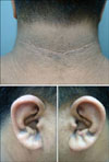
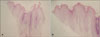
A 23-year-old man presented with brown-black plaques on the posterior neck, axillae, groin, and both auricles. The hyperpigmented plaques began to develop one year prior to arrival, and since that time plaques on the neck and both auricles had slowly darkened. Physical examination showed symmetric distribution of hyperpigmented velvety brown plaques on the posterior neck, axillae, and groin. Several brown-black plaques were visible along the sulci of the pinnae (Fig. 1). The patient weighed 120 kg, with a BMI of 39. He had been diagnosed with fatty liver three years prior, but otherwise he denied any significant medical or family history. Routine laboratory evaluations, including complete blood count and urinalysis, showed normal findings. Blood chemistry revealed elevated AST (52 U/L, normal range 8~40 U/L), ALT (149 U/L, normal range 5~40 U/L), and triglycerides (308 mg/dl, normal range 34~143 mg/dl). Plaques on the posterior neck, axillae, and groin were consistent with obesity-associated acanthosis nigricans. The clinical differential diagnoses of the auricular lesions included verruca plana, epidermal nevus, and seborrheic keratosis. A biopsy specimen from the auricle revealed papillomatosis, hyperkeratosis, and focal parakeratosis in the epidermis, as well as some spores on the stratum corneum. Neither acanthosis nor koilocytes were observed, however, and no inflammatory cell infiltration was found in the dermis (Fig. 2). On the basis of these clinical and histological findings, the auricular lesion was diagnosed as acanthosis nigricans.
DISCUSSION
Schwartz3 classified 8 types of acanthosis nigricans: benign acanthosis nigricans, obesity-associated acanthosis nigricans, syndromic acanthosis nigricans, malignant acanthosis nigricans, acral acanthosis nigricans, unilateral acanthosis nigricans, medication-induced acanthosis nigricans, and mixed-type acanthosis nigricans. Among these, obesity-associated acanthosis nigricans is the most common. Other than the patient's obesity, no pathologic finding to explain the cause of acanthosis nigricans could be found on physical and laboratory examinations. Topical and retinoid creams were prescribed for the skin lesions, which have shown some gradual improvement.
Acanthosis nigricans is commonly observed in the flexural areas, including the neck, axillae, and groin. Rarely, acanthosis nigricans involves mucosal surfaces, such as the oral cavity. We could not find a previous report of acanthosis nigricans on the auricle associated with obesity. Herein, we report a rare and interesting case of obesity-associated acanthosis nigricans involving the pinnae of the ears.
 XML Download