

 PDF
PDF ePub
ePub Citation
 Print
Print


INTRODUCTION
Dermal melanocytosis is characterized by the presence of ectopic melanocytes in the dermis. The most common forms include the Mongolian spot, blue nevus, nevus of Ota, and nevus of Ito. Some cases of dermal melanocytosis do not fit into any of these morphologic categories, however. Dermal melanocytosis rarely occurs in unusual regions or covers extensive areas of the body1. Atypical types of dermal melanocytosis reported in the literature include dermal melanocyte hamartoma and congenital segmental dermal melanocytosis2,3.
We herein report the clinical and histological features of an unusual case of congenital dermal melanocytosis.
CASE REPORT
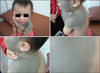
A 21-month-old Korean girl presented with an asymptomatic pigmented patch on her left face, neck, chest, shoulder, and back that had been present since birth. The pigmented patch had grown proportionately to the patient's size throughout her life; otherwise, there had been no appreciable change in its color or texture. The patient had no family history of pigment disorders, and her physical and mental development was normal. On physical examination, the patient had an extensive amount of uniform deep blue patches that covered the left side of her face, neck, chest, shoulder, and back in a unilateral pattern, with a few dark blue-brown macules in the lesions.
There were also scattered satellite lesions of a similar appearance on her backbone area (Fig. 1). The epidermal surface was unaltered, and no underlying induration was clinically apparent. Except for the skin lesions, there were no remarkable findings on physical examination. There was no discoloration or other abnormality on ophthalmologic examination.
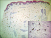
A biopsy specimen from a lesion on the patient's back demonstrated a number of elongated melanocytes located throughout the dermis. Melanocytes were numerous in the upper and middle dermis. Immunohistochemical analysis showed that melanocytes and melanin were positive for S-100 and Fontana-Masson stains, but cells were negative for CD68, which can be a marker for melanophages. Melanocytes were scattered among the collagen bundles, and some of these cells were aggregated around blood vessels. Their elongated cytoplasms were loaded with fine melanin (Fig. 2). Nevus cell nests were not seen in the epidermis or dermis. Appendages were normal.
DISCUSSION
Except for blue nevi, the various types of dermal melanocytosis are similar in their histopathologic characteristics; they just differ in the concentration and location of the melanocytes. The characteristics they share include elongated dermal melanocytes that are widely scattered between collagen fibers in the dermis. These melanocytes do not alter the normal topography of the skin3.
In our case, the skin lesion involved the face and acromioclavicular region, respectively sites for the nevus of Ota and the nevus of Ito, but our patient's lesion extensively covered the chin and neck with no involvement of the eye. In nevi of Ota and Ito, the melanocytes are mainly situated in the upper dermis, and the basal layer of the epidermis may be hyperpigmented. However, in our case, melanocytes were also found in the middle and lower dermis.
Typical Mongolian spots occur in the sacrococcygeal region as pale-blue to slate-gray macular hyperpigmentation. Occasionally, however, they occur outside the lumbosacral region on places such as the middle or upper part of the back as aberrant Mongolian spots. Histopathologically, the melanocytes of Mongolian spots predominantly reside in the lower portions of the dermis. These features are clinically and histopathologically different from our case.
The blue nevus usually presents as a well-demarcated, blue-black papule, nodule or plaque that is generally acquired. Histologic features of the blue nevus include a high concentration of dermal melanocytes in the middle and lower third of the dermis. Melanophages and variable degrees of fibrosis are present. These features are different from our case.
A few cases of unusual congenital dermal melanocytosis have been documented (Table 1). In 1981, Bashiti et al.1 reported a female infant with generalized blue-gray discoloration of the skin. In the same year, Burkhart and Gohara2 described an 18-month-old male with diffuse, bilateral gray-blue pigmentation on his buttocks that extended in a dermatomal pattern down the entire length of his right leg. They used the term "dermal melanocyte hamartoma" to describe this phenomenon. In 1992, Vélez et al.3 documented the case of a 28-year-old white woman with extensive, speckled, gray-blue pigmentation in a segmental pattern on the right side of her trunk. They proposed the term "congenital segmental dermal melanocytosis" to describe the findings. In 1999, Grézard et al.4 reported a 45-year-old Caucasian woman with a slowly spreading bilateral congenital pigmentation of the back in a dermatomal distribution from the fourth to the eighth dorsal dermatome that extended laterally to the external part of the breasts.
Several authors have also documented cases of isolated small patch forms of congenital dermal melanocytosis5-7. The pathogenesis of congenital dermal melanocytosis is not well understood. It is believed that during embryogenesis, dermal melanocytes migrate from the neural crest to their designated site at the epidermal-dermal junction. Dermal melanocytosis occurs when the melanocytes fail to properly reach that designated site8.
Our case has some similarities to those of Burkhart and Gohara2 and Vélez et al.3, but the unilateral distribution and melanocytes throughout the dermis did not fit into any of the cases that have been reported. As a result, we propose the term, "congenital unilateral dermal melanocytosis," to name the lesion on our patient.
We herein report a case of congenital unilateral dermal melanocytosis.
 XML Download