

 PDF
PDF ePub
ePub Citation
 Print
Print


INTRODUCTION
Sarcoidosis is a multisystemic disease that is defined by the formation of noncaseating granulomas in different organs. The patients who have systemic sarcoidosis may develop granulomatous infiltration of the subcutaneous tissue as a specific cutaneous lesion, and this is referred to as subcutaneous sarcoidosis1. The most frequent specific cutaneous lesions of sarcoidosis are lupus pernio, infiltrated plaques, maculopapular eruptions, infiltration of old scars and subcutaneous sarcoidosis2,3. Subcutaneous sarcoidosis is the least frequent of these specific cutaneous lesions. It has been reported to occur in 1.4% to 6% of patients who have systemic sarcoidosis4.
CASE REPORT
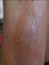
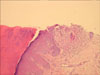
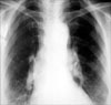
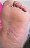
A 53-year-old female patient was admitted with a painless swelling on her left foot, and she'd had this swelling for the previous eight months. She was otherwise asymptomatic. There was no history of scar or trauma. On physical examination, an erythematous subcutaneous nodule 3 cm in size was detected at the plantar surface of the left foot (Fig. 1). Magnetic resonance imaging revealed two irregular shaped solid lesions that were 3×2 cm and 1×2 cm in size at the left foot plantar aponeurosis. Tru-cut biopsy of the lesion showed non-necrotizing granulomatous inflammation (Fig. 2, 3). A chest radiograph revealed bilateral hilar enlargement with bilateral reticulonodular opacities (Fig. 4) and thorax computed tomography showed multiple mediastinal and bilateral hilar lymphadenopathies and bilateral extensive milimetric nodules at both lung fields. No endobronchial lesion was observed during flexible bronchoscopy, and the CD4/CD8 ratio in the bronchoalveolar lavage fluid was 2.9. Transbronchial needle aspiration from a subcarinal lymph node was performed and pathological examination revealed noncaseating granulomatous inflammation. The serum angiotensin-converting enzyme (ACE) level was 78 UI/L (N: 8~52 UI/L). The pulmonary function tests were normal. The radiologic staging of sarcoidosis is made according to chest radiography and it is as follows; (0) normal, (I) bilateral hilar lymphadenopathy, (II) bilateral hilar lymphadenopathy and parenchymal infiltrates, (III) parenchymal infiltrates without bilateral hilar lymphadenopathy and (IV) signs of fibrosis5. Based on these findings the diagnosis of stage II sarcoidosis and subcutaneous sarcoidosis was made. Systemic therapy was not administered since the patient was asymptomatic, the pulmonary function tests were normal and any other organ involvement was not detected. However, for the subcutaneous lesion, two intralesional triamcinolone acetonide injections with a dose of 10 mg/ml was applied one month apart. Complete resolution was observed after the second application (Fig. 5).
DISCUSSION
Subcutaneous sarcoidosis was first described by Darier and Roussy in 1904 as a rare cutaneous form of sarcoidosis4,6. Although several cases of subcutaneous sarcoidosis have been reported, this entity is thought to be rare. However, a review of the Korean dermatologic literature showed that subcutaneous nodules are the most common form of cutaneous sarcoidosis in Korean patients7.
Most of the reported cases of subcutaneous sarcoidosis have occurred in Caucasian women and most often in the fourth and fifth decades, like in our patient. The lesions are characteristically located on the extremities and they are usually bilateral and asymmetric1. Other sites such as the trunk, face, head and neck have also been reported. However, plantar involvement is very rare and to the best of our knowledge there has been only one such report in the literature8. The nodules are frequently firm, painless, mobile, round and without discoloration9. The range of the number of lesions is from 1 to 100, and the average size of these lesions is 0.5 to 2 cm. Several authors have emphasized that the subcutaneous nodules tend to be fusiform10-12.
In most of the reported cases and in our presented case subcutaneous sarcoidosis is associated with systemic sarcoidosis and especially bilateral hilar adenopathy. Although subcutaneous sarcoidosis has been considered to more frequently appear late in the course of the disease, the recent reports suggest that it usually appears at the beginning of sarcoidosis10,13-15. Bilateral hilar lymphadenopathy with or without mediastinal adenopathy is by the far the most common chest involvement9. In the largest series by Ahmed and Harstad (a total of 21 patients), a systemic disease component was recognized in 16 of the 20 patients who underwent a systemic evaluation at the time the subcutaneous sarcoidosis was diagnosed13. They reported that all 16 of these patients had pulmonary involvement, which was documented by chest radiography. Bilateral hilar adenopathy was identified in 15 of the 16 patients (94%); in 6 of these cases, paratracheal adenopathy and pulmonary infiltrates were also present and pulmonary infiltrates were identified in the one patient with no hilar adenopathy. The lesions were characteristically located on the extremities and they were usually bilateral and asymmetric (the upper extremity in 21 patients, the lower extremity in 16, the trunk in 6, the buttocks in 2 and the forehead in 1). In 20 of the 21 patients (95%), more than one anatomic site was involved, and all 20 of these patients had lesions on an upper extremity. They also reviewed the literature and identified 33 cases with a diagnosis of subcutaneous sarcoidosis. They reported that the extremities were involved in 26 patients (78.8%) and this included the upper and lower extremities in 14, the upper extremity in 7 and the lower extremity in 5. More than one anatomic site was involved in 19 patients (58%). A systemic disease component was present in 29 patients (88%) at the time the diagnosis of subcutaneous sarcoidosis was established. Choi et al.16 reported on a case with the nodular type of subcutaneous sarcoidosis and the patient had a nontender, palpable soft tissue mass on the left buttock.
When systemic sarcoidosis is suspected, the search for specific cutaneous or subcutaneous lesions is especially important. Because subcutaneous sarcoidosis usually appears at the beginning of the disease, the demonstration of sarcoid granulomas in subcutaneous tissue may be useful for establishing the diagnosis of systemic sarcoidosis and to avoid more aggressive diagnostic procedures. On the other hand, the development of firm, painless nodules that are mainly located on the forearms in otherwise asymptomatic patients obligates one to rule out systemic sarcoidosis.
The treatment of cutaneous sarcoidosis is often difficult with a high rate of recurrence. While non-specific lesions are often associated with a good prognosis, specific lesions are more chronic and they tend to resolve with scarring. Topical corticosteroids may be effective. Kang et al.17 reported a successful outcome with using topical corticosteroid in a patient with cutaneous sarcoidosis that presented as multiple erythematous macules and patches. The severity of the systemic disease determines the need and modality of treatment. For patients with severe systemic involvement or disfiguring skin lesions, the mainstay of treatment is systemic corticosteroid therapy. In patients who cannot tolerate systemic corticosteroids, the use of alternative therapies, including methotrexate and hydroxychloroquine, has been reported to be helpful18. However, there is still not a standard treatment regimen for subcutaneous sarcoidosis. In patients with only a few nodules, treatment with intralesional corticosteroids has also been attempted, but with limited success18. Fichtel et al.8 gave minocycline 100 mg orally twice a day and 15 drops of a saturated solution of potassium iodide and they observed a complete resolution after 3 months of treatment. In contrast, in our case, complete improvement was achieved with intralesional corticosteroid treatment.
In conclusion, although it is very rare, sarcoidosis must be considered for a patient who presents with subcutaneous nodules at the plantar surface of the foot, and a systemic evaluation should also be considered. Our patient achieved a successful outcome after intralesional corticosteroid treatment.

 XML Download