

 PDF
PDF ePub
ePub Citation
 Print
Print


INTRODUCTION
Lichen planus pigmentosus (LPP) was first described by Bhutani et al.1, and this a rare variant of lichen planus, and LPP appears as mottled or reticulated hyperpigmented, dark brown macules or papules on the sun exposed areas and flexural folds2,3. Linear lichen planus, which shows a linear distribution following Blaschko's lines, is considered a subtype of lichen planus3-5.
CASE REPORT
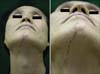
A 60-year-old man presented with an asymptomatic hyperpigmented linear patch on the anterior neck and chin and he'd had this patch for 8 month. One dark brown macule first appeared on the neck, and it gradually spread to the chin. There was no history of excessive sun exposure or trauma on that site. On examination, he had a linear streak of dark brown patches from the chin and across the neck following one of Blaschko's lines (Fig. 1). There was no other skin lesion and the oral mucosa and nails were not involved. He had previously suffered with gastric cancer and he had undergone gastrectomy at the age of 47 years.
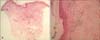
The routine laboratory data, including the peripheral blood cells and liver function tests, was normal. Skin biopsy showed epidermal thinning, basal cell degeneration, some dyskeratotic cells with pigment incontinence and a lymphohistiocytic infiltration in the dermis (Fig. 2). He was treated with systemic and topical steroid, but the lesion still remained unchanged for 7 months.
DISCUSSION
LPP is characterized by mottled or reticulated hyperpigmented, dark brown macules on the sun exposure skin areas and flexural folds on the face and neck, and the axillae and inguinal regions1-3.
The cause of LPP is unknown, but an immunologic mechanism mediates its development, as well as that of lichen planus4. Based on the distinctive lymphocytic inflammatory response of the lichenoid reactions, cell mediated immunity seems to play a pivotal role in triggering the clinical expression of the disease. T-lymphocytes are pivotal as they regulate epidermal cell recognition, the lichenoid response and epithelial destruction2-4,8. Clinically, LPP differs from classical lichen planus by exhibiting dark brown macules and/or papules and a longer clinical course2.
Kanwar et al.6 described flexural involvement over the axillae (8.9%), inflamed skin folds (6.5%) and groin (3.2%) in 123 Indian patients with LPP. The histopathological findings of LPP consist of hyperkeratosis, atrophic epidermis with vacuolar alteration of the basal layer and scarce lymphohistiocytic or lichenoid infiltrates in the dermis with pigment incontinence1-3,8.
Although there have been a few reports of LPP, there have been only 2 reports of LPP with a linear pattern in the Korean dermatologic literature2. Our patient had lesion on the neck and chin with a linear pattern. The linearity of the lesion is probably related to Blaschko's lines, which suggests that the predisposition to develop LPP might be determined during embryogenesis5.
Blaschko's lines are thought to reflect T-lymphocytic migration and the clonal expression during embryogenesis of the skin. The genetic mosaicism in an acquired Blaschko-linear inflammatory dermatosis could be responsible for the cutaneous antigenic mosaicism that may induce a mosaic T-cell response according to the trigger of viral infection or drugs. Some apoptotic change in our cases may be responsible for the response of the mosaic T cells that are present along Blaschko's lines2,5.
The differential diagnosis of our case includes linear dermatosis such as lichen striatus, linear and whorled nevoid hypermelanosis, ashy dermatosis and postinflammatory hyperpigmentation. Lichen striatus almost always has preceding inflammatory papules or a scaly eruption, which last for 4 months to 4 years and a perivascular and periadnexal inflammatory cell infiltration4,8. There were no previous papules on our patient and the histological findings were more consistent with LPP. Linear and whorled nevoid hypermelanosis usually presents within a few weeks of birth and it is associated with congenital anomalies. The histologic findings have shown basal pigmentation without pigment incontinence3,4. In ashy dermatosis and postinflammatory hyperpigmentation, the histopathologic findings show a slightly smoothed epidermis with a scanty perivascular lymphocytic infiltration. However, there was a dense lymphohistiocytic infiltration in our case. Although erythema multiforme and fixed drug eruptions show histopathological findings similar to those of our case, there was no apparent epidermal necrosis in our case.
In conclusion, LPP can present along the lines of Blaschko. Therefore, we should consider LPP in the differential diagnosis of a linear pigmentary disorder.
 XML Download