Abstract
Sarcoidosis is an idiopathic multisystem disease with various cutaneous presentations, and it is characterized by the presence of non-caseating granulomas in the affected organs. The specific manifestations are papules, plaques, nodules, ulcers and scar. We report here on a variant of sarcoidosis on a 71-year-old woman who showed an indurated plaque on the forearm. Her lesion's appearance was clinically similar to that of a morphea and the appearance of the lesion was unlike the commonly observed manifestations of sarcoidosis.
Sarcoidosis is a multisystem disease that is histopathologically characterized by the presence of non-caseating granulomas in the affected organs. Cutaneous lesion occurs in 20~35% of the patients with systemic sarcoidosis. Because of the wide variety of skin manifestations of sarcoidosis, a classification has been proposed that divides them into specific and nonspecific lesions. The specific cutaneous lesions of sarcoidosis are characterized by non-caseating granulomas, and the clinical picture of the specific skin lesions is highly heterogeneous as a wide variety of different cutaneous manifestations have been reported1,2. Here we report on a variant of sarcoidosis that was clinically similar to a morphea.
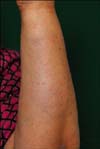
A 71-year-old woman had a 1 month history of a firm plaque on her left forearm, which had started as an erythematous patch and it had gradually became more indurated. The lesion was asymptomatic and there was no history of previous trauma to the affected area. She had hypertension and diabetes, and these illnesses were being treated with losartan, thiazide, metformin and nateglinide. Physical examination showed a whitish indurated plaque on the left forearm and no tenderness (Fig. 1). A biopsy specimen showed sarcoidal granuloma in the lower dermis and subcutaneous fat tissue, asteroid bodies, prominent dermal sclerosis and highly situated eccrine glands (Fig. 2). The polarized light examination did not reveal any foreign bodies. Special stains for acid fast bacilli and fungi were negative. The polymerase chain reaction test for mycobacteria was also negative.
A diagnosis of morpheaform sarcoidosis was made based on the clinical and histopathological findings. The routine laboratory investigations did not find significant abnormalities. The anti-nuclear, anti-SSA, anti-SSB and anti-SCL-70 antibodies were all negative. The purified protein derivative test was also negative. Her chest X-ray findings were normal. However, the result of the pulmonary function test showed a restrictive pattern. Furthermore, increased airway hyperreactivity in response to methacholine was observed. The patient was treated with triamcinolone intralesional injection. Within 2 months, the induration had considerably improved.
The diagnosis of cutaneous sarcoidosis is made based on the clinical manifestations, the histopathologic examination and the laboratory findings. Sarcoidosis remains a diagnosis of exclusion. Even a skin biopsy is not sufficient for making the diagnosis of sarcoidosis3. Therefore, making the differential diagnosis with other possibilities, such as tuberculosis, atypical mycobacterial infection, fungal infection or a foreign body reaction, is essential4. Our patient did not have any systemic abnormalities except for the pulmonary function test. However, a normal chest X-ray may be observed in 5 to 15% of the patients with sarcoidosis. A restrictive pattern of lung impairment and increased airway hyperreactivity are also common manifestations of sarcoidosis5.
Morpheaform sarcoidosis is an uncommon manifestation of cutaneous sarcoidosis and to the best of our knowledge only 5 cases have been previously described in the English medical literature. Of these 5 previously reported patients, 3 were black and 2 were white. The clinical features are indistinguishable from those of true morphea, and the cutaneous lesions may precede or arise years after the extracutaneous sarcoidosis. Only 1 patient had a cutaneous sarcoidosis without any other systemic involvement and it is remarkable that 3 of the patients had chronic arthritis, which is a rare manifestation of sarcoidosis. Histopathologically, morpheaform sarcoidosis is characterized by a prominent dermal sclerosis, which is correlated with the clinical induration6-8. The mechanism of sclerosis has not yet been determined, yet several cytokines released by the activated T-cells and macrophages from a granuloma could be the stimuli for dermal sclerosis9.
The treatment of cutaneous sarcoidosis is often difficult with a high rate of recurrence. New therapeutic options have been introduced, including pentoxifylline, infliximab and etanercept. However, the worldwide accepted standard therapies include the administration of corticosteroids, antimalarials and methotrexate10. The treatment of morpheaform sarcoidosis in the previous cases was similar to the standard therapies6-8. An intralesional steroid injection was effective for our patient.
In conclusion, we clinically experienced a case of morpheaform sarcoidosis on the arm of a 71-year-old woman, and this might be the first such reported case in Korea. Physicians should consider sarcoidosis in the differential diagnosis of indurated skin lesions.
Figures and Tables
References
1. Kerdel FA, Moschella SL. Sarcoidosis. An updated review. J Am Acad Dermatol. 1984. 11:1–19.
2. Elgart ML. Cutaneous sarcoidosis: definitions and types of lesions. Clin Dermatol. 1986. 4:35–45.

3. Costabel U, Guzman J, Baughman RP. Systemic evaluation of a potential cutaneous sarcoidosis patient. Clin Dermatol. 2007. 25:303–311.

4. Kang MJ, Kim HS, Kim HO, Park YM. Cutaneous sarcoidosis presenting as multiple erythematous macules and patches. Ann Dermatol. 2009. 21:168–170.

5. Lynch JP 3rd, Ma YL, Koss MN, White ES. Pulmonary sarcoidosis. Semin Respir Crit Care Med. 2007. 28:53–74.

6. Hess SP, Agudelo CA, White WL, Jorizzo JL. Ichthyosiform and morpheaform sarcoidosis. Clin Exp Rheumatol. 1990. 8:171–175.
7. Burov EA, Kantor GR, Isaac M. Morpheaform sarcoidosis: report of three cases. J Am Acad Dermatol. 1998. 39:345–348.


 PDF
PDF ePub
ePub Citation
 Print
Print


 XML Download