

 PDF
PDF ePub
ePub Citation
 Print
Print


INTRODUCTION
Lupus vulgaris is the most common form of cutaneous extension of tuberculosis disease from underlying affected tissue such as bone, joints or lymphoid tissue, or from hematogeneous or lymphatic spread of mycobacteria from an endogenous focus in a previously sensitized host with a high level of cell-mediated immunity1. Misdiagnosis and delayed treatment of lupus vulgaris sometimes occur because of the variations in the clinical presentation and the low rate of positive cultures2. We report here on a case of a longstanding and uncommon annular form of lupus vulgaris that masqueraded as tinea cruris.
CASE REPORT
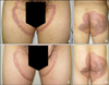
A 36-year-old man presented with well-defined, progressive, asymptomatic, annular skin lesions on his inguinal areas and buttocks for 10 years. He had previously received many different therapies such as antifungal, antibiotic and corticosteroid therapies for these lesions. Especially, he had been treated with oral and topical antifungal agents for several years under the diagnosis of tinea cruris at several clinics, and none of these medications was effective. On physical examination, we observed well demarcated, irregular bordered, violaceous, annular indurated margins on his groin and buttocks (Fig. 1A, B).
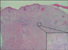
The results of the laboratory tests, including a complete blood cell count, a basic biochemical profile and chest X-ray, were within normal limits. A 4-mm punch biopsy specimen was obtained from the affected area for microscopic examination. The biopsy specimen showed acanthosis and irregular papillomatosis of the epidermis. The dermis revealed frequent non-caseating epithelioid granulomas and a heavy lymphohistiocytic infiltration (Fig. 2). Acid-fast bacilli were not found on the Ziehl- Neelsen staining. A 2TU purified protein derivative (PPD) test was positive with 20 mm indurations after 48 hours. A polymerase chain reaction (PCR) assay revealed the presence of Mycobacterium tuberculosis DNA in a lesional biopsy specimen.
A diagnosis of lupus vulgaris was made based on the cutaneous symptoms, and this was supported by the histopathological findings and positive PCR assay. He received a four-drug protocol (isoniazid 300 mg/day, rifampicin 600 mg/day, ethambutol 1,000 mg/day, pyrazinamide 1,500 mg/day) for the first 2 months, followed by isoniazid and rifampicin maintenance therapy for another 7 months. The cutaneous lesions started to regress within 3 months and they healed with atrophic scarring at 9 months (Fig. 1C, D).
DISCUSSION
Lupus vulgaris is usually a re-infection of tuberculosis of the skin and this is acquired either exogenously by direct inoculation of the bacilli or endogenously by hematogenous or lymphatic spread from an underlying infected focus1. In some cases, lupus vulgaris may develop exogenously following inoculation for tuberculosis secondary to Bacille Calmette-Guerin vaccination3.
This malady classically presents as a solitary asymptomatic plaque with an atrophic centre and an infiltrated, serpiginous or polycyclic red-brown border. The plaque is formed by coalescence of soft, friable, gelatinous papules4. In European populations, 80% of the lesions are localized to the head and neck region, followed by the arms and legs4,5. In Turkey, lupus vulgaris has a predilection for the face (62%), followed by the forearms, chest, trunk and legs6. Misdiagnosis of lupus vulgaris sometimes occurs because of its rarity or its sporadic presentation in atypical forms7,8. In our case the annular lesions were localized on the thighs and buttocks, which is an uncommon form and site for lupus vulgaris. Our patient had also been misdiagnosed as suffering from tinea cruris or eczema by a local practitioner, and antifungal and steroid treatment had given no relief for a long time. The lesions had been improperly treated for about 10 years until our diagnosis of lupus vulgaris was made.
Histopathological studies show tubercules or tuberculoid granulomas with slight or absent caseation in the papillary dermis and a variable degree of epidermal hyperplasia9,10. The histopathological differential diagnosis includes sarcoidosis, tuberculoid leprosy, granulomatous foreign body reactions and so on. Making the histological and clinical differentiation of tuberculoid leprosy from lupus vulgaris is very difficult, if not impossible7,8. So not only histopathological examination, but also mycobacterial culture, a PPD test and PCR are important methods for diagnosing cutaneous tuberculosis. The microbial culture of lupus vulgaris is frequently negative, and only a 6% positivity rate has been reported for the cutaneous cultures from patients with lupus vulgaris.
The management of lupus vulgaris is similar to that for tuberculosis of other organs. But the localized forms of lupus vulgaris that are without evidence of associated internal tuberculosis may be treated with isoniazid alone for up to 12 months. A total dose of 80 to 140 g isoniazid may be required. Because viable mycobacteria have been found in clinically healed lesions, the treatment should be continued for at least 2 months after complete involution of the lesions11.
We report here on a case of lupus vulgaris that had uncommon clinical manifestations, and these manifestations mimicked fungal infection. We think that the diagnosis of lupus vulgaris should be kept in mind for patients with long standing skin lesions that do not respond to the routine treatments.
 XML Download