

 PDF
PDF ePub
ePub Citation
 Print
Print


INTRODUCTION
Heterotopic gastric mucosa has the histologic features of gastric mucosa but is situated outside the boundaries of the stomach, anywhere from the mouth to the rectum1. There have also been reported cases seen in other areas of the digestive system: in intestinal duplications, Meckel's diverticulum, the gallbladder or cystic duct, the omphalomesenteric duct and the liver2-9. Occurrences in the umbilicus, however, are an extremely rare and peculiar phenomena. Following an extensive literature search, we found only 1 case of heterotopic gastric mucosa in the umbilicus.
We report this case of heterotopic gastric mucosa in the umbilical area and our thoughts regarding the cause of this phenomenon.
CASE REPORT
A 5-month-old male patient presented with a pink, erythematous, slowly enlarging, slimy nodule on the umbilicus, present since birth. The infant was born following an uneventful gestational period and had no congenital anomalies.
The umbilical cord dried and fell off at 10 days, however, an erythematous nodule was observed on the umbilical base. Topical electrical cauterization had been performed several times, though the mass continued to grow.
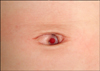
Physical examination showed a dome-shaped, bright red papule with a 1-cm diameter in the umbilical area (Fig. 1). Neither bleeding nor a foul smell was noted.
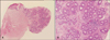
With a presumed diagnosis of an umbilical granuloma or urachal duct remnant, the patient had received abdominal computed tomography which demonstrated no anomaly. We therefore performed a wide excision of the lesion. Microscopic examination of the excised skin lesion showed a polypoid lesion which was composed of well-developed chief cells, parietal cells, and dilated mucous glands (Fig. 2). Heterotopic gastric mucosa was diagnosed. The patient had no symptoms for 1 year following the operation.
DISCUSSION
Umbilical nodules are not rare in infants and young children. The most common cause is umbilical granuloma, which are nodules that vary from benign lesions to severe congenital anomalies. Radiological imaging and a biopsy are therefore needed10.
"Heterotopia," derived from Greek, refers to the finding of normal tissue in a foreign site entirely separate from the main organ. Heterotopic gastric mucosa have been observed throughout the alimentary tract, from the oral cavity to the rectum. They have also been seen in intestinal duplications, Meckel's diverticulum, the gallbladder or cystic duct, the omphalomesenteric duct and the liver2-4. In addition to the alimentary tract, occurrences in the urinary bladder, the spinal column, the salivary glands, bronchogenic cyst, and the thyroglossal cyst, as well as in intra-abdominal and intra-thoracic locations, have all been noted in literature7-9.
Despite an extensive literature search, we found only one other case of gastric heterotopia located in or near the umbilicus. This previous case was heterotopic gastric mucosa and pancreatic tissue in the skin of the abdominal wall5.
While there is no clear explanation for the cause of heterotopic tissue growth, it is possible that these growths arise from heteroplastic differentiation, developmental accidents, and metaplastic differentiation11. In the gallbladder, it is thought that the fundic-type gastric mucosa occurs as a result of either a developmental anomaly or heteroplastic differentiation, while most pyloric-type gastric mucosa occur following metaplastic differentiation12. In the head and neck region, a possible mechanism for gastric heterotopia may be developmental errors related to the proximity of the gastric anlage and other fore-gut structures, resulting in the isolation of the gastric mucosa during its morphologic migration13.
We postulate the following occurred in our patient: In the developmental process, the mid-gut elongates and herniates into the umbilical cord during the 6th week of embryogenesis. Within the umbilical cord, the mid-gut rotates 90° counterclockwise around the axis of the superior mesenteric artery, while at the same time elongating to form the jejunum and ileum, and the lumen of the omphalomesenteric duct closes. By the 10th week of embryogenesis, the mid-gut returns to the abdominal cavity14. It is at this point in the process when gastric mucosa cells could be seeded in the umbilical area, giving rise to heterotopic gastric mucosa.
We herein report a very rare case of heterotopic gastric mucosa of the skin as well as a possible cause of this phenomenon.
 XML Download