

 PDF
PDF ePub
ePub Citation
 Print
Print


INTRODUCTION
Linear focal elastosis, also known as elastotic striae, is clinically characterized by asymptomatic, palpable, striae, which extend horizontally across the middle and lower back. Histopathologically it can be seen as having deposition of abnormal elastic fibers in the dermis1-3. Until now, only 2 cases of linear focal elastosis with a family history have been reported4,5. Here, we present one such case, of linear focal elastosis with a family history.
CASE REPORT
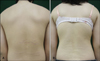
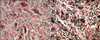
A 15-year-old Korean boy presented with erythematous linear depressed patches on his back that had developed 2 months prior to his visit (Fig. 1A). His 13-year-old sister presented with whitish linear palpable plaques on her back, which had been present for 2 years (Fig. 1B). The depressed patches and palpable plaques were asymptomatic and extended horizontally. Neither patients had a recent history of rapid or excessive weight gain or loss, no history of medications, and no trauma to their backs. The skin biopsy specimen taken from the older brother showed normal epidermis with thin, fragmented abnormal elastic fibers in the dermis. As well, the number of elastic fibers was decreased (Fig. 2A). The specimen taken from the younger sister showed increased deposition of thin, fragmented abnormal fibers in the dermis (Fig. 2B). In both patients, the clinical and histopathological findings were consistent with the diagnosis of linear focal elastosis. They were treated with topical retinoids for several months, however there was no marked improvement.
DISCUSSION
Linear focal elastosis is an uncommon elastotic disorder. Clinically, it is characterized by asymptomatic, palpable, striae, which extend horizontally across the middle and lower back. Histopathologically it is distinguished by deposition of abnormal elastic fibers Which separate the dermal collagen bundles1-3. There has been no effective treatment thus far for linear focal elastosis.
The pathophysiology of linear focal elastosis is unclear. Some believe that it could be the result of an extreme degenerative/regenerative process6. Early lesions appear erythematous, are more active and proliferate, and histopathologically show elastolysis, whereas older lesions lack erythema, appear more static, and show elastogenesis6. This pattern is suggestive of a degenerative/regenerative process that results in the eventual formation of elastotic lesions. In our case, the older brother presented with a 2-month history of erythematous linear depressed patches on his back. Microscopic examination showed a deposition of abnormal, fine, fragmented elastic fibers which are decreased in numbers. The younger sister presented with a 2-year history of whitish linear palpable plaques on her back. Microscopic examination showed massive, thin, fragmented abnormal elastic fibers that are increased in numbers. These findings are compatible with early and old lesions of linear focal elastosis, respectively.
Clinical differential diagnosis of linear focal elastosis may include striae distensae, anetoderma and pseudoxanthoma elasticum. Striae distensae are characterized by atrophic linear bands and a wrinkled appearance, and are usually located on the abdomen, thighs, arms, or breast. They are associated with steroid use, pregnancy, and weight change3. The histopathology of striae distensae shows a flattened epidermis, atypical collagen fibers, and variable changes in elastic tissue1. However, in our case, both patient shows horizontally oriented atrophic or palpable striae-like lines on their back, without any medication or weight change. Histopathologic findings demonstrated normal epidermis with thin, fragmented abnormal elastic fibers in the dermis but no changes in the collagen fibers. The histological presence of elastosis is a distinguishing feature from anetoderma, which presents with a loss of elastic fibers. Pseudoxanthoma may be differentiated by the presence of calcified elastic fibers.
Approximately 20 cases of linear focal elastosis have been described. To our knowledge, only two cases of linear focal elastosis having a family history have been reported. Moiin and Hashimoto4 reported the case of a black man whose father had had similar lesions since childhood, and Kim et al.5 reported a case of twins who had similar asymptomatic yellowish palpable lesions on the lower back.
Here, we add another case, making it three, by describing a rare familial case of linear focal elastosis with typical features, which occurred in a brother and sister.
 XML Download