

 PDF
PDF ePub
ePub Citation
 Print
Print


INTRODUCTION
Kikuchi's disease (KD) is a benign, usually self-limited disease characterized by cervical lymphadenopathy and fever1,2. Other symptoms may include leucopenia, liver dysfunction, weight loss, diarrhea, nausea, vomiting, myalgia, and arthralgia3,4. Less frequently, KD involves skin lesions as erythematous macules, papules, patches, and plaques on the scalp, chest, back and extremities5,6. We report a 20-year-old Korean woman with KD that showed unique skin involvement with symmetrically distributed, erythematous, firm nodules limited to the face.
CASE REPORT
A 20-year-old Korean woman was referred to Department of Dermatology from Department of Internal Medicine for multiple erythematous hard nodules on the face. She was initially admitted to the Department of Infectious Disease due to remitting fever, chills, and persistent and painful cervical lymphadenopathy. She reported a 3 week history of fever, malaise, and progressive cervical lymph node enlargement, and a 1 week history of erythematous macules on both cheeks. Prior to coming to our hospital, the patient was managed conservatively with oral antibiotics, non steroidal anti-inflammatory drugs, and topical clindamycin. This treatment failed to improve general symptoms and skin lesions.
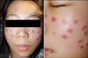
On physical examination, the patient was febrile (37.9℃) and pale. The posterior cervical lymph nodes ranged in diameter from 1 to 2 cm and were palpable on the right side of the patient's neck. Cutaneous examination showed asymptomatic multiple erythematous firm nodules distributed symmetrically on both cheeks (Fig. 1). A skin biopsy showed patchy infiltration of mononuclear cells around the skin appendages and blood vessels. The infiltrating cells were composed of large, atypical lymphocytes and histiocytes with crescent-shaped nuclei and many apoptotic cells. On immunohistochemical staining, the lymphoid cells were positive for CD3 but negative for CD20 or CD56. Some cells were also positive for granyzme B, and histiocytic cells were positive for CD68 (Fig. 2).
Laboratory testing showed leucopenia (white blood cell count 1.7×10⁹/L) and thrombocytopenia (platelet count 136×109/L). The erythrocyte sedimentation rate was 36 mm in the first hour. Liver and renal function tests, urinalysis, and chest radiograph showed no abnormalities. Serum complement (C3, C4) levels were normal and the serology for venereal disease research laboratory test, rheumatoid arthritis factor, antinuclear antibody, double-stranded DNA antibodies, anti-Ro and anti-La antibodies were negative. A lymph node biopsy showed mixed infiltrates of histiocytes, plasmacytoid monocytes, and lymphoid cells with necrosis, typical features of KD.
The patient was treated with prednisolone 20 mg/day for 2 weeks. She became afebrile, and the lymphadenopathy and cutaneous lesions regressed. Prednisolone was gradually tapered over the next 2 weeks. She remained asymptomatic over a 4-month follow-up period.
DISCUSSION
KD is a rare, self-limited lymphadenopathy first reported in 1972 as a distinct clinicopathological entity of unknown etiology1,2. It mainly affects young adults, mostly women, and is clinically characterized by cervical lymphadenopathy, high fever, occasional leucopenia, and liver dysfunction. Other symptoms may include weight loss, diarrhea, nausea, vomiting, thoracic pain, myalgia, arthralgia, and skin rashes3,4.
Apoptosis may play an important role in KD pathogenesis. In patients with KD, cytotoxic T lymphocytes may act as apoptotic effectors as well as target cells, while histiocytes may enhance apoptosis7,8. A viral pathogenesis, including Epstein-Barr, human herpesvirus, HIV and parvovirus B19, may cause KD because of the self-limited clinical course of this disease and the lack of a neutrophilic response9-11.
KD is associated with autoimmune disorders, mainly systemic lupus erythematosus (SLE), mixed connective tissue disease, anti-phospholipid syndrome, thyroiditis, polymyositis, and scleroderma5,6,12. The strongest link seems to be with SLE, although the exact nature of this association has not yet been established. Diagnosis is made by histopathological examination of lymph node biopsy tissues4. Histologically, the lymph nodes exhibit focal, well-circumscribed, paracortical, necrotic foci, surrounded by histiocytes, immunoblasts, and plasmacytoid monocytes and the absence of neutrophils13.
Many patients with KD have no cutaneous findings, but skin eruptions occur in up to 40% of cases, including urticarial, morbilliform, rubella-like or drug-eruption-like rashes, generalized erythema and papules, plaques and nodules, leukocytoclastic vasculitis, erythema multiform, papulopustules, and even eyelid edema5,6,11. These lesions are usually located on the trunk, upper extremities, or the whole body, including the face6,12. This patient showed a unique clinical appearance, with cutaneous manifestations presenting as multiple hard erythematous nodules distributed symmetrically on the face only.
Skin biopsies of KD patients have demonstrated variable histopathological findings, including nonspecific superficial and deep perivascular infiltrates, papillary dermal edema and vacuolar changes with necrotic keratinocytes at the dermal-epidermal junction, as well as patchy infiltrates of lymphoid cells, histiocytes and nuclear debris, and findings similar to those observed in lymph node biopsies. Dermal infiltrates consisting of histiocytic cells, so called plasmacytoid monocytes, and karyorrhectic debris, were positive for CD686,10,14.
KD usually resolves spontaneously without any treatment in 2~3 months, although it can recur14. Corticosteroids used for short periods in patients with serious clinical symptoms help to speed up the resolution process. The cutaneous eruption of KD resolves in a few weeks to months, and the course is similar to that of a lymphadenopathy12.
In conclusion, the cutaneous clinical findings of KD are often nonspecific. In a patient with skin findings (including erythematous nodules distributed symmetrically on the face), fever, and lymphadenopathy, KD should be considered in the differential diagnosis.
 XML Download