

 PDF
PDF ePub
ePub Citation
 Print
Print


INTRODUCTION
Neurofibromatosis is one of the most common autosomal dominant inherited disorders. In addition to multiple skin manifestations, patients with neurofibromatosis have an increased risk for benign or malignant tumors. Eccrine spiradenoma, first described by Kersting and Helwig in 1956, is a benign tumor of the sweat glands. It presents as a painful, slow-growing and solitary nodule on the head or upper trunk. Neurofibromatosis with eccrine spiradenoma has not been reported in English literature. Here, we report a rare case of eccrine spiradenoma in a patient with neurofibromatosis.
CASE REPORT
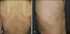
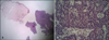
A 50-year-old male presented in our clinic with the complaint of a 30-year-old painful skin-colored subcutaneous mass on his back. He had axillary freckles and numerous café-au-lait macules and soft nodules on his trunk and limbs. One of these soft nodules was biopsied and determined to be a neurofibroma by histological examination. He had no other significant health problems, and his father, brother, sister and son were diagnosed with neurofibromatosis. Physical examination revealed a 4 cm-sized, skin-colored tender round subcutaneous mass on his back (Fig. 1). Laboratory examinations, including blood and urine tests and chest X-ray, were within normal limits. Histopathological examination of the tender mass revealed well-demarcated lobules in the dermis without connections to the epidermis. The epithelial cells within the tumor lobule were arranged in intertwining bands. Two types of epithelial cells were seen in the bands. The cells of the first type had small, dark nuclei located at the periphery of the bands. The cells of the second type had large, pale nuclei located in the center of the bands. Diffuse lymphocytic infiltrations in the tumor lobule were also observed (Fig. 2). Immunohistochemistry revealed that the tumor cells were stained positively for cytokeratin. The histopathologic findings were consistent with eccrine spiradenoma and treated with total excision of the tumor. Now he is under observation without recurrence.
DISCUSSION
Neurofibromatosis is an autosomal dominant inherited disorder. Riccardi et al, in 1986, classified this disorder into 8 types according to clinical manifestations. But currently, it is classified as neurofibromatosis type 1 (NF-1) and neurofibromatosis type 2 (NF-2) based on the known genes involved. NF-1, also known as Von Recklinghausen's disease, is the most common neurofibromatosis that accounts for about 85% of all cases. NF-1 is due to a depletion of the NF-1 gene encoded in chromosome 17; whereas, NF-2 results from a depletion of the NF-2 gene on chromosome 22. NF-1 affects approximately 1 in 3,000~5,000 people, across all ethnic groups. Along with skin findings such as café-au-lait macules, multiple neurofibroma and axillary freckles, many disorders of the neural, skeletal, endocrine and vascular system can coexist1.
Patients with NF-1 are at a 2~5 times increased risk for developing neoplasms compared with normal population2. These neoplasms include CNS tumors such as optic nerve glioma, acoustic neuroma, meningiomatosis, neuroblastoma and also pheochromocytoma, thyroid carcinoma3. A relationship among juvenile xanthogranuloma, neurofibromatosis and juvenile chronic myeloid leukemia is strongly suggested. Patients with neurofibromatosis accompanied by juvenile xanthogranuloma showed a 2~30 times increased risk for leukemia than those with neurofibromatosis alone. Therefore, informing patients with both neurofibromatosis and juvenile xanthogranuloma about the risk is necessary4-6. Other benign tumors such as granular cell and glomus tumors can also accompany neurofibromatosis7,8.
A relationship with malignant neoplasms of skin, including malignant melanoma, basal cell carcinoma, Merkel cell carcinoma and malignant nodular hidradenoma, is also suggested2,9. In the case of malignant melanoma, there is strong relevance to neurofibromatosis, because both conditions originate from the neural crest, and many cases have been reported10-12.
Eccrine spiradenoma is a benign uncommon neoplasm originating from the eccrine gland. Clinically, it presents as a painful, slow-growing, solitary nodule on the head or upper trunk. It occurs in young and middle-aged adults, without predilection for either gender. It is usually solitary; but rarely, it presents as numerous nodules in a zosteriform pattern or linear arrangement. Histopathologically, eccrine spiradenoma presents as one or more intradermal lobules surrounded by a fibrous capsule without connections to the epidermis. On higher magnification, the epithelial cells within the tumor lobule are arranged in intertwining cords. Two types of epithelial cells are present in the cords. The cells of the first type have small, dark nuclei located at the periphery of the cellular aggregates. The cells of the second type have large, pale nuclei arranged around a small lumina13,14.
To date, no case of neurofibromatosis accompanied by an eccrine spiradenoma has been reported in the English literature, so it is uncertain whether eccrine spiradenoma coexists incidentally or results from a genetic defect associated with neurofibromatosis, like other tumors. If neurofibromatosis has a genetic connection with eccrine spiradenoma, these diseases would occur together more frequently. Because those 2 diseases have a different origin, and reported cases are rare, this case is likely to be incidental in nature.
We report a rare case of eccrine spiradenoma occurring in a patient with neurofibromatosis, which has never been reported in the English literature.
 XML Download