

 PDF
PDF ePub
ePub Citation
 Print
Print


INTRODUCTION
Sweet syndrome (SS) is an acute febrile neutrophilic dermatosis that is clinically characterized by fever, neutrophilia and painful erythematous skin lesions, and it is histologically characterized by a predominantly mature neutrophilic dermal infiltrate1. Neutrophilic dermatosis of the dorsal hands (NDDH) is considered to be a rare localized variant of SS with less frequent systemic symptoms. NDDH begins on the hands and it can spread to other locations2. We present here the case of a 34-year-old woman with recurrent solitary tender lesions on the right hand and lips, but she had no associated systemic symptoms.
CASE REPORT
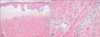
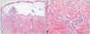
A 34-year-old woman presented with a one-week history of erythematous skin rash on the hand. This lesion had recurred each spring for the last four years. She had another cutaneous lesion on the right lips that had first developed one day previously. Her medical and family histories were noncontributory. She was receiving no medications. She had no underlying diseases such as hematologic malignancy or inflammatory bowel disease. Physical examination revealed a solitary, tender, erythematous, well-demarcated plaque on the dorsum of the right thumb (Fig. 1A) and an erythematous plaque with small vesicles on her right lips (Fig. 1B). She had a febrile and chilling sensation one week previously. She had no fever, arthralgia or generalized malaise at the time of diagnosis. Biopsy specimens from both the lesions on the dorsal hand and lips revealed edema of the papillary dermis and a dense perivascular infiltrate of neutrophils with leukocytoclasia throughout the upper to mid dermis, but there was no evidence of true vasculitis (Fig. 2, 3). Gram staining of the tissue was negative. The laboratory studies revealed a normal erythrocyte sedimentation rate (ESR). Her white blood cell count was 9,800 cells µl-1 with 68.8% neutrophils, the haemoglobin level was 13.5 g/dl and the platelet count was 330,000/µl. The result of the serum electrolyte assessment, the renal and liver function panel and the urinalysis were normal. The serum albumin and alkaline phosphatase were normal.
A diagnosis of NDDH was made, and the patient was treated with prednisone 20 mg daily for one week and then 5 mg daily for the following one week. The lesions resolved without scarring or recurring during the 6 months of follow-up.
DISCUSSION
NDDH is a rare, localized variant of SS, and this was first described by Galaria et al.3 in 2000. The pathogenesis of SS may be multifactorial and it remains to be definitively determined4. A septic process, a hypersensitivity reaction, leukotactic mechanisms and cytokines have all been postulated to contribute to the pathogenesis of SS4. Clinically, NDDH is characterized by tender, erythematous plaques, pustules and bullae that are generally limited to the dorsal hands and fingers3. This is usually limited to the dorsal hands, and often with a predilection for the lateral aspect of the hand between the thumb and index finger5. However, only several cases have been reported in which there were concurrent lesions located on either the arm, leg, back and/or face1.
The histology of NDDH shows prominent papillary dermal edema and a dense diffuse infiltration of mature neutrophils throughout the upper dermis3. Swollen endothelial cells, dilated small blood vessels and fragmented neutrophil nuclei can also be present4. Fibrin deposition or neutrophils within the vessel walls (the changes of "primary" leukocytoclastic vasculitis) is usually absent4. In 1995, Strutton et al.5 described six patients with hand lesions that clinically resembled NDDH, but the lesions had the histological features of leukocytoclastic vasculitis, and so he proposed a new entity called "pustular vasculitis of the hands". However, Gilaberte et al.6 suggested that the vasculitis might be a secondary event related to the intensity of the neutrophilic infiltrate and the time of evolution of the lesions.
The clinical presentation of NDDH differs from that of classic SS. In classic SS, fever, leukocytosis and an increased ESR are observed in 80~90% of the cases; inflammatory bowel disease (16%) and hematologic disease (54%) may be present as the accompanying diseases4,7. In NDDH, the lesions are clinically restricted to the hands; fever, leukocytosis and an increased ESR are observed in 33% of the cases and hematologic malignancy is present as an accompanying disease in 21% of the cases7.
The differential diagnosis of NDDH includes allergic contact dermatitis, cellulitis and pyoderma gangrenosum (PG). Allergic contact dermatitis is an eczematous dermatitis that presents as severe pruritus that develops in regions exposed to allergen, and allergic contact dermatitis shows spongiotic dermatitis histologically8. Cellulitis or erysipelas is excluded according to the negative tissue cultures and stains and a lack of response to antibiotics3. Atypical pyoderma gangrenosum, also known as vesiculobullous PG, presents with chronic hemorrhagic bullous lesions that ulcerate superficially and are usually seen in patients with leukemia or polycythemia vera7. However, pyoderma gangrenosum may be histologically indistinguishable from NDDH, although the first usually shows true vasculitis7. Thus, atypical PG and pustular vasculitis of the dorsal hands are arguably within the spectrum of a single disease entity, which is most appropriately termed NDDH9.
The treatment for NDDH is believed to be same as that for SS. Many different treatments have been used for SS with different rates of success and relapse10. These include corticosteroids, dapsone, potassium iodide, colchicine, clofazimine, azathioprine, danazol, tetracyclines and cyclosporine. Systemic corticosteroids are the most common first-line therapy10. Walling et al.7 showed that treatment with systemic corticosteroids was successful in 71% of the cases.
In summary, we have presented a case of recurrent NDDH with no associated systemic signs and symptoms and the patient was successfully managed with low dose systemic corticosteroids. There has been only one case of NDDH with bacterial endocarditis in the dermatological literature11. The remarkable characteristic of our patient was the concomitant involvement of the lips. We suggest that dermatologists consider this disease when they encounter a patient with a tender erythematous bullous lesion occurred on the dorsal hands.

 XML Download