

 PDF
PDF ePub
ePub Citation
 Print
Print


INTRODUCTION
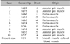
Smooth muscle hamartomas (SMH) are a result of benign proliferation of smooth muscle bundles in the dermis. SMH-associated smooth muscle cells originate from arrector pili-, dartos-, vulvar-, mammillary- and vascular wall-muscles1-3. SMH is characterized by slightly pigmented plaques that contain vellus hairs, and SMH is subdivided into two types: congenital smooth muscle hamartoma (CSMH) and acquired smooth muscle hamartoma (ASMH)1-3. ASMH is a very rare form of SMH that was first described by Wong and Solomon1 in 1985, with only such 10 cases having been reported in the English medical literature (Table 1). These lesions usually occur on the proximal extremities1,2, scrotum4-7, vulva8, shoulders2 and trunk9-11.
CASE REPORT
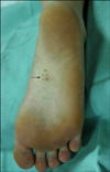
A 21 year-old women presented with an 18-month history of a slow-growing 0.5×0.5 cm nodule on the right sole (Fig. 1). She complained that the lesion was mildly tender and somewhat numb. Any evidence of trauma, allergic reaction, hyperpigmentation and hypertrichosis was not found and the Pseudo-Darier's sign could not be elicited. Moreover, the results of the complete blood count, blood chemistry and urinalysis were all within the normal limits.
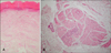
A skin biopsy specimen from the lesion revealed bundles of smooth muscle cells with cigar shaped nuclei, and these cells were arranged haphazardly throughout the dermis and especially around the blood vessels (Fig. 2). Furthermore, the spindle cells stained positive with Masson trichrome stain and α-smooth-muscle actin antibodies. The nodule was subsequently excised, and there has been no evidence of recurrence.
DISCUSSION
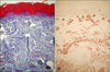
SMH is usually manifested at birth, but it can also appear later in life. CSMH is a congenital lesion that's principally composed of smooth muscle with or without vellus hair and/or epidermal hyperpigmentation. It is usually located on the trunk and extremities and it appears as variable sized papules, patches or plaques4. Transient piloerection or elevation of a lesion that's induced by rubbing is referred to as Pseudo-Darier's sign and this is the characteristic diagnostic clue for CSMH1-4. ASMH appears later in life as a slightly hyperpigmented or hypertrichotic patch or plaque with associated itching, pain or numbness. These lesions are usually negative for Pseudo-Darier's sign7. The histopathologic hallmark of SMH is a local increase of large, randomly oriented bundles of smooth muscle cells with central, cigar-shaped nuclei. These smooth muscle bundles are arranged haphazardly around blood vessels in the dermis, and the smooth muscle bundles stain positive with Masson trichrome stain and α-smooth-muscle actin antibodies (Fig. 3).
The differential diagnosis for SMH includes leiomyomas, angioleiomyomas and Becker's nevus. In contrast to SMH, the smooth muscle bundles in leiomyomas are more tightly interlacing and poorly demarcated, and they can also be painful or tender. Angioleiomyomas contain thick-walled blood vessels with smooth muscle fibers deposited in concentric layers that merge with the muscular stroma. Hence, angioleiomyomas can be differentiated from SMH by the histological findings.
Becker's nevus often resembles SMH both clinically and histologically. Becker's nevus is a well-defined, uniformly hyperpigmented plaque with hypertrichosis. Histologically, Becker's nevus exhibits an abnormal number of large, deeply seated terminal hair follicles that are sometimes associated with hyperplastic pilar smooth muscles. However, Becker's nevus can also present with a proliferation of dermal smooth muscle. Because of the overlapping histological features, some authors have speculated that Becker's nevus and SMH are two polar entities at either end of a continuous spectrum2,4,9. Since Wong and Solomon1 reported on the first case of ASMH in 1983, only 10 cases of ASMH have currently been reported in the English medical literature. ASMH is usually located on the skin of the forearm, chest, neck, scrotum, penis, major labium or shoulder. Five cases of ASMH had been reported to originate from the dartos muscle and 5 cases have been reported to originate from the arrector pili muscle (Table 1). In this case, we hypothesize that the hamartomatous proliferation of smooth muscle cells originated from blood vessel walls due to the haphazard arrangement of smooth muscle bundles around the dermal vasculature and the fact that arrector pili and dartos muscles are not normally present in foot soles. To the best of our knowledge, this is the first case of a smooth muscle hamartoma of the sole to be reported in the English medical literature.
 XML Download