Journal List > Ann Dermatol > v.21(1) > 1045179

Haw and Lee: A Case of Giant Folliculosebaceous Cystic Hamartoma

Abstract

Clinically, folliculosebaceous cystic hamartoma (FSCH) lacks distinct features, but it has been reported as an asymptomatic, 1- to 3-cm, dome-shaped nodule on the face. Histopathologically, FSCH is characterized by adnexal and folliculosebaceous cystic proliferation with various mesenchymal changes. This case presented an unusually large, small-fist-sized mass in the right mandibular area that was accompanied by intermittent itching. Histopathologic findings demonstrated appropriate features of FSCH. We present an interesting case of giant FSCH in a 48-year-old female.

INTRODUCTION

Kimura et al1 originally reported eruptive folliculosebaceous cystic hamartoma (FSCH) in 1991. Clinically, FSCH is usually a solitary, 0.5- to 1.5-cm, flesh-colored, sessile or pedunculated papule on the head, especially on the central part of the face and nose2.
Because few cases of giant FSCH have been reported, there is no distinct definition of "giant". According to previous reports3-6, these lesions range from 5 cm to 23 cm in diameter. Along with its larger size, and unlike ordinary FSCH, giant FSCH occurs on areas other than the face.

CASE REPORT

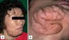
A 48-year-old female presented with a pruritic skin-colored soft tumor on her right lower cheek that had appeared as small papules 20 years earlier and had been growing gradually. The skin lesion was a skin-colored, cerebriform mass, sized 10×8×5 cm, on the right mandibular area (Fig. 1A). Its smooth surface contained scattered, dilated hair follicle-like pores with a waxy discharge (Fig. 1B).
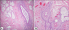
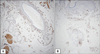
Histological examination showed numerous sebaceous lobules radiating from cystic follicular structures with mesenchymal changes. Numerous mature sebaceous lobules surrounded the cystic structures. The cystic structures were lined with stratified squamous epithelium and showed well-developed, dilated infundibular portions of hair follicles with infundibular keratinization. There were some increases of muscle components near the sebaceous lobules (Fig. 2A), but no secondary hair follicles. The folliculo-cystic structures were wrapped in densely laminated collagen bundles with various vascular proliferation (Fig. 2B). In immunohistochemical staining, muscle components were immunoreactive to desmin and smooth muscle actin (Fig. 3A). Increased vascular structures showed reactivity to CD31 (Fig. 3B), but tumor stromal cells were negative for S-100 protein and neurofilament.
The size of the lesion decreased slightly after treatment with isotretinoin (30 mg/d) for 3 months, but returned to its original size after cessation of drug. We recommended that the patient undergo excision and skin grafting but she did not want further treatment.

DISCUSSION

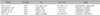
FSCH is a distinct cutaneous hamartoma formed from epithelial and stromal elements. Among the cases of FSCH in the literature1-20, only a few cases of giant FSCH have been reported (Table 1)3-6. The clinical features of our case were similar to those of the reported giant FSCH cases, except for cerebriform surface and chin involvement. FSCH is slightly more common in females. The sites of predilection are the nose, cheeks, forehead, and scalp. In most cases, the lesion does not exceed 3 cm in diameter7. The four cases in the literature described as "giant" ranged from 5 cm to 23 cm in diameter. The lesions in giant FSCH cases occurred in various sites, such as genital areas, upper extremities, and upper back.
FSCH has distinct, unique, classical histopathologic features. Kimura et al1 established the histological criteria for diagnosis of FSCH as: 1. an infundibular cystic structure attached to sebaceous lobules via sebaceous ducts; 2. laminated fibroplasias around the entire fibroepithelial units; 3. mesenchymal changes around fibroepithelial units; 4. clefts between fibroepithelial units and surrounding altered stroma; 5. confinement of the process primarily to the dermis. Several variations in FSCH reflect the predominance of either epithelial or non-epithelial components. Fogt and Tahan8 reported large expanses of tufted aggregates, separated or enveloped within the epithelial units. Donati and Balus9 presented a case with neural proliferation, while Aloi et al10 described copious mucin deposits in a fibrillary stroma. Our case involved several mesenchymal changes, such as laminated collagen, vascular proliferation, and slightly increased muscle components.
Because of its peculiar histopathologic features, diagnosis of FSCH is not difficult. But, in some cases, FSCH must be differentiated from sebaceous trichofolliculoma. Sebaceous trichofolliculoma is associated with pilosebaceous hyperplasia and overgrowth5. It presents as a central cystic cavity, usually containing a hair shaft, with sebaceous lobules and secondary hair follicles. However, sebaceous trichofolliculoma has minimal surrounding stromal proliferation. In FSCH, there are no secondary hair follicles and hair shaft in the cystic cavity. Mesenchymal change in FSCH is another point of differential diagnosis.
Giant FSCH is less common than ordinary FSCH and this particular case occurred on the face unlike other giant FSCH cases. We need to gather more cases to determine how FSCH and giant FSCH differ from each other in clinical features and prognosis.

Figures and Tables

Fig. 1
(A) The skin lesion is a skin-colored, cerebriform mass, 10.0×8.0×5.0 cm, in the right mandibular area. (B) The smooth surface has scattered, dilated hair follicle-like pores with a waxy discharge.
ad-21-63-g001
Fig. 2
(A) Numerous mature sebaceous lobules surround the cystic structures. There is some increase of muscle components on the left lower quadrant (H&E, ×40). (B) Increased numbers of small venules are shown around the fibroepithelial units (H&E, ×100).
ad-21-63-g002
Fig. 3
(A) A slight increase of smooth muscles is shown around fibroepithelial units (SMA, ×40). (B) Increased numbers of small venules are shown around fibroepithelial units. Some of these vessels are congested with red blood cells (CD31, ×40).
ad-21-63-g003
Table 1
The clinical features of the reported cases of giant folliculosebaceous cystic hamartoma
ad-21-63-i001

References

1. Kimura T, Miyazawa H, Aoyagi T, Ackerman AB. Folliculosebaceous cystic hamartoma. A distinctive malformation of the skin. Am J Dermatopathol. 1991. 13:213–220.
2. Templeton SF. Folliculosebaceous cystic hamartoma: a clinical pathologic study. J Am Acad Dermatol. 1996. 34:77–81.
crossref
3. Sturtz DE, Smith DJ, Calderon MS, Fullen DR. Giant folliculosebaceous cystic hamartoma of the upper extremity. J Cutan Pathol. 2004. 31:287–290.
crossref
4. Yamamoto O, Suenaga Y, Bhawan J. Giant folliculosebaceous cystic hamartoma. J Cutan Pathol. 1994. 21:170–172.
crossref
5. Hamada M, Kiryu H, Satoh E, Moroi Y, Urabe K, Furue M. A case of genital folliculosebaceous cystic hamartoma with an unique aggregated manifestation. J Dermatol. 2006. 33:191–195.
crossref
6. Brücher JJ, Franke I, Ulrich J, Gollnick H, Leverkus M. Giant genital variant of folliculosebaceous cystic hamartoma: successful management by CO2 laser and acitretin therapy. Br J Dermatol. 2007. 157:833–835.
crossref
7. El-Darouty MA, Marzouk SA, Abdel-Halim MR, El-Komy MH, Mashaly HM. Folliculo-sebaceous cystic hamartoma. Int J Dermatol. 2001. 40:454–457.
crossref
8. Fogt F, Tahan SR. Cutaneous hamartoma of adnexa and mesenchyme. A variant of folliculosebaceous cystic hamartoma with vascular-mesenchymal overgrowth. Am J Dermatopathol. 1993. 15:73–76.
crossref
9. Donati P, Balus L. Folliculosebaceous cystic hamartoma. Reported case with a neural component. Am J Dermatopathol. 1993. 15:277–279.
10. Aloi F, Tomasini C, Pippione M. Folliculosebaceous cystic hamartoma with perifollicular mucinosis. Am J Dermatopathol. 1996. 18:58–62.
crossref
11. Yamamoto T, Ohkubo H, Nishioka K. Folliculosebaceous cystic hamartoma associated with rosacea. J Dermatol. 1993. 20:712–714.
crossref
12. Donati P, Balus L, Bellomo P. Folliculosebaceous cystic hamartoma. J Am Acad Dermatol. 1997. 36:502.
crossref
13. Toyoda M, Morohashi M. Folliculosebaceous cystic hamartoma with a neural component: an immunohistochemical study. J Dermatol. 1997. 24:451–457.
crossref
14. Ramdial PK, Chrystal V, Madaree A. Folliculosebaceous cystic hamartoma. Pathology. 1998. 30:212–214.
crossref
15. Bolognia JL, Longley BJ. Genital variant of folliculosebaceous cystic hamartoma. Dermatology. 1998. 197:258–260.
crossref
16. Schulz T, Hartschuh W. Folliculo-sebaceous cystic hamartoma is a trichofolliculoma at its very late stage. J Cutan Pathol. 1998. 25:354–364.
crossref
17. Simon RS, de Eusebio E, Alvarez-Vieitez A, Sanchez Yus E. Folliculo-sebaceous cystic hamartoma is but the sebaceous end of tricho-sebo-folliculoma spectrum. J Cutan Pathol. 1999. 26:109.
crossref
18. Zimmermann T, Hartschuh W, Raulin C. Facial folliculosebaceous cystic hamartoma. Treatment with CO2 and Er:YAG lasers. Hautarzt. 2004. 55:289–291.
19. Brasanac D, Boricic I. Giant nevus lipomatosus superficialis with multiple folliculosebaceous cystic hamartomas and dermoid cysts. J Eur Acad Dermatol Venereol. 2005. 19:84–86.
crossref
20. Yamagata K, Mitsuishi T, Kawana S. Folliculosebaceous cystic hamartoma with hair shaft fragments. Eur J Dermatol. 2005. 15:105–106.
TOOLS
Similar articles