

 PDF
PDF ePub
ePub Citation
 Print
Print


INTRODUCTION
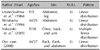
Ashy dermatosis is a peculiar, slowly progressive, idiopathic dermal melanosis that was first described by Ramirez in 19571. Clinically, ashy dermatosis consists of slate gray- to lead-colored patches and ranging in size from 3 mm to very large confluent patches, surrounded by an erythematous, slightly elevated border2,3. In most cases, the trunk, arms, neck, and face are involved symmetrically2. Only three cases of unilateral ashy dermatosis have been reported (Table 1)4-6.
CASE REPORT
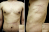
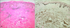
A 27-year-old Korean man presented to our clinic with progressive cutaneous pigmentary changes on his left back, flank, and abdomen. One year earlier, erythematous patches appeared on his left back and spread gradually to the left flank and abdomen. After 3 months, portions of the erythematous patches became hyperpigmented. The patient complained only of intermittent itching. He was on no medications and there was no family history of hyperpigmentation. The physical examination revealed hyperpigmented patches surrounded by erythematous patches in a linear distribution along Blaschko's line (Fig. 1). Laboratory tests, including a complete blood count, blood chemistry analysis, urinalysis, and VDRL, were within the normal range or negative. Histopathologic findings showed hyperpigmentation and vacuolar degeneration along the basal layer of the epidermis. They also showed pigment incontinence and perivascular lymphocytic infiltration in the dermis (Fig. 2A). Fontana-Masson stains showed increased epidermal melanin and some pigment incontinence (Fig. 2B).
DISCUSSION
Ashy dermatosis is a chronic, idiopathic dermal melanosis characterized by hyperpigmented macules. The patches of variable shape and size are an ashen-gray color and primarily involve the face, neck, trunk, and proximal arms2,3. The patient can be of any age, but the mean age of occurrence is in the 2nd decade of life2,7. Ashy dermatosis is most common in Asia and Central and South America, and is somewhat more common in women2,3,7. The etiology of ashy dermatosis is unknown. Ashy dermatosis has been reported in association with the ingestion of ammonium nitrate, intestinal parasites (whipworm infection), orally administered contrast medium, cobalt allergy in plumbers, vitiligo, human immunodeficiency virus (HIV) infection, and chronic hepatitis C2,3,7.
Clinically, the lesions of ashy dermatosis vary in color from slate gray to lead, and in size from 3 mm to very large confluent patches2. Individual lesions can be oval, irregular, or even polycyclic in shape, and continue to grow slowly. In most cases, the lesions are surrounded by an erythematous, non-scaling, slightly elevated border3,6. The trunk, arms, neck, and face are commonly involved symmetrically, although there are three reports of ashy dermatosis with a unilateral distribution (Table 1)4-6. Yokozeki et al.6 described unilateral ashy dermatosis distributed along Blaschko's lines in the first reported case in 2005. Another two cases have been reported in the Japanese literature4,5. Our case is the second case of unilateral ashy dermatosis in the English literature.
The differential diagnosis of unilateral ashy dermatosis includes lichen planus pigmentosus (LPP), partial unilateral lentiginosis (PUL), and post-inflammatory hyperpigmentation (PIH)6,7. LPP consists of brown-to-black patches without an erythematous border on the face and flexor folds. PUL usually presents during childhood and has no erythematous border. The histopathologic findings of prominent pigment incontinence in PIH and late stage ashy dermatosis are hard to differentiate; however, PIH can be ruled out clinically by a history of preceding cutaneous insults, such as drug or phototoxic reactions, infections, physical injury or trauma, allergic reactions, and inflammatory diseases.
Histopathologically, ashy dermatosis may show basal vacuolar changes in the epidermis, superficial perivascular lymphohistiocytic infiltration, and pigment incontinence3. A recent study reported a predominance of HLA-DR+, ICAM-1+ keratinocytes and CD8+ T lymphocytes8.
There is no definitive treatment for ashy dermatosis. Recently, clofazimine has been used with some success in adults, apparently because of its anti-inflammatory and immunomodulating effects9. A successful response to dapsone in one patient has also been reported10.
We have reported a rare case of unilateral ashy dermatosis with a linear distribution related to Blaschko's lines on the left abdomen, flank, and back. In addition, we suggest the possibility that ethnic and geographic factors are involved in the prevalence of unilateral ashy dermatosis.
 XML Download