

 PDF
PDF ePub
ePub Citation
 Print
Print


INTRODUCTION
Lichen planus (LP) is a unique, common inflammatory disorder that affects the skin, mucous membranes, nails, and hairs. LP is characterized by pruritic, violaceous papules that are most commonly situated on the extremities of middle-aged adults1.
Palmoplantar LP, an acral, localized variant is rare and may create difficulty in diagnosis if it is present as an isolated finding. Highly pruriginous, erythematous, scaly plaques with or without hyperkeratosis, and located on the internal plantar arch without involvement of the fingertips are characteristic of palmoplantar LP. The lesions resemble psoriasis vulgaris, warts, calluses, porokeratosis, hyperkeratotic eczema, tinea, or secondary syphilis2.
We report a rare case of a 37-year-old male with LP of which multiple erythematous, scaly, hyperkeratotic papules were limited to the palms and soles, creating a misdiagnosis of secondary syphilis.
CASE REPORT
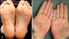
A 37-year-old male presented with erythematous, scaly papules on his palms and soles of 3 months duration. He had transferred to our outpatient clinic from a local medical center with the clinical impression of secondary syphilis. The patient did not complain of any subjective symptoms, including an itching sensation, nor did he have any unusual sexual contact history. On physical examination, no lymphadenopathy was noted. On the skin examination, multiple rice grain- to bean-sized, erythematous, scaly, hyperkeratotic papules and hyperpigmented macules were observed on both internal plantar arches and palms symmetrically; the fingertips were spared (Fig. 1). The other skin surfaces, including hair, nails, and oral mucosa, were normal. The VDRL, TPHA, and FTA-ABS were negative.
Histopathologically, marked hyperkeratosis, focal parakeratosis, hypergranulosis, and irregular acanthosis with a saw-tooth like appearance on the epidermis were observed. Vacuolar changes of the basal layer and band-like lymphohistiocytic infiltration on the upper dermis were also found (Fig. 2).
After the diagnosis of palmoplantar LP was established, he was given topical steroid ointment (0.05% clobetasol-17-propionate) twice a day for 3 months. Four months later, the lesions had improved.
DISCUSSION
In 1869 the term lichen planus (Greek leichen, "tree moss"; Latin planus, "flat") was coined by Eramus Wilson3. The exact incidence and prevalence of LP are unknown; LP occurs throughout the world in all races. A pruritic, papular eruption of LP is characterized by its violaceous color, polygonal shape, and sometimes, fine scales. LP is most commonly found on the flexor surfaces of the wrists, arms, and legs, the genitalia, and mucous membranes2. According to the site of involvement, LP is categorized as LP of the scalp and nails, inverse LP, and palmoplantar LP2.
Palmoplantar LP is a clinical variant of LP and usually involves the palms and/or soles, as well as other sites of the skin. However, LP limited to the palms and/or soles is very rare and has been reported in only 2 cases in the Korean dermatologic literature. One case with involved LP limited to the soles in a 50-year-old female who presented with erythematous, hyperkeratotic-crusted, pruritic plaques on both soles4. The other case was LP in a 26-year-old male who presented with erythematous, hyperkeratotic, pruritic papules on his palms and soles5. The latter case was misdiagnosed as secondary syphilis and treated with benzathine penicillin-G with no effect, as in our case. Both cases presented with characteristic pruritus, but our 26-year-old male patient did not complain of any subjective symptoms. According to Sanchez-Perez et al.6, palmoplantar involvement in LP is not uncommon and the lesions mostly begin outside the palmoplantar region; however, none of the cases of LP were limited to the palms and soles. Typical LP is more common in women and most patients are in their 30s to 60s, whereas palmoplantar LP is more common in men and in their 20s to 40s6. Highly pruriginous, erythematous, scaly plaques with or without hyperkeratosis are characteristic. The lesions are most commonly found on the internal plantar arch and yellow, compact, keratotic papules or papulonodules are seen on the lateral margins of the fingers and hand surfaces; however, they are less likely to affect the fingertips6.
Palmoplantar LP may share cutaneous findings among psoriasis vulgaris, warts, calluses, tinea, or secondary syphilis. Therefore, histopathologic examination is essential for the differential diagnosis. Palmoplantar LP presents the same histopathologic findings as classic LP, which is characterized by basal epidermal keratinocyte damage and lichenoid-interface lymphocytic reaction, such as hyperkeratosis, wedge-shaped hypergranulosis, irregular acanthosis, necrotic keratosis (Civatte body), Max-Joseph space, dense band-like lymphocytic infiltration in the papillary dermis, and vacuolar alteration of basal layer2,6,7. Although parakeratosis is thought not to be a feature of LP of the skin7, Sanchez-Perez et al.6 reported that focal parakeratosis was observed in 17 of 38 biopies obtained from the palmoplantar region in 36 patients with LP, and we also observed focal parakeratosis in our case.
The cutaneous findings in secondary syphilis are characterized by symmetric, usually polymorphous, and less often scaly, maculopapular rashes with a copper hue and sharply demarcated margins on the face, shoulders, flanks, and anal or genital regions. Unlike LP, secondary syphilis usually involves eruptions on the palms and soles8. Interestingly, there are two case reports in the PubMed database, which were secondary syphilis presenting as a LP-like rash distributed over the upper arm8, or over the forehead, upper back, dorsum of the feet, and palms9. The histopathologic findings of the both mimicked a lichenoid drug eruption.
In conclusion, this interesting case of LP with erythematous, scaly, hyperkeratotic papules limited to the palms and soles which created a misdiagnosis of secondary syphilis emphasizes to clinicians that cutaneous lesions of LP are varied and may be limited to the palms and soles, and that histopathologic examination is essential for the appropriate differential diagnosis.

 XML Download