

 PDF
PDF ePub
ePub Citation
 Print
Print


INTRODUCTION
Herpes zoster is caused by the varicella zoster virus. Following natural infection, the virus remains latent in the sensory dorsal root ganglion cells and replicates at some later time. The clinical manifestations are characterized by several groups of painful vesicles on an erythematous and edematous base situated unilaterally within the distribution of a cranial or spinal sensory nerve1. Bilateral involvement and recurrence are rare. Zoster, even more rarely, involves two widely separated regions at one time2. The phenomenon of zoster occurring in two noncontiguous, widely separated dermatomes has been referred to as zoster duplex unilateralis or bilateralis3. In Korea, four cases of zoster duplex bilateralis have been reported4-7.
We report a rare case of herpes zoster involving noncontiguous dual dermatomes on both halves of the body.
CASE REPORT
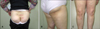
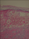
A 67-year-old woman presented with a seven day history of pain on her right buttock and thigh. Two days after onset of the pain, grouped, vesicular eruptions developed along the right buttock, and posterior aspect of the thigh and knee (dermatome L4~L5) (Fig. 1). Five days after the onset of pain, the same symptoms developed along the left anterior chest and left side of the back (dermatome T7~T8) (Fig. 2). The patient was acutely ill-looking in appearance; the history was significant for hypertension, osteoporosis, and a lumbar spine compression fracture. Laboratory studies included a complete blood cell count, routine urinalysis, liver function tests, VDRL, and a chest PA. All results were negative or within normal limits. A Tzanck smear and skin biopsies were performed on the vesicles of the right buttock. Multinucleated giant cells with intranuclear inclusion bodies were observed on the Tzanck smear. The skin biopsy showed vesicles, ballooning degeneration, and multinuclear giant cells in the epidermis (Fig. 3). The patient was treated with oral administration of famcyclovir 750 mg/day for seven days and daily wet dressings until all vesicles had crusted over. The skin lesions and pain subsided without complications.
DISCUSSION
Herpes zoster (HZ) is characterized by the reactivation of a latent varicella zoster virus (VZV) residing in the dorsal root ganglion after primary infection3. Herpes zoster is a relatively common disease (10% to 20% lifetime risk of HZ) and manifests as several groups of painful vesicles with a characteristic distribution along the dermatomes that is usually unilateral. The dermatomes most frequently affected are in the thoracic region2. The skin lesions of HZ are classically limited to a single dermatome. Non contiguous HZ involving multiple dermatomes is very rare in both immunocompetent and immunocompromised persons. When zoster occurs in two non contiguous dermatomes, it is referred to as zoster duplex unilateralis or bilateralis, depending on whether one half or both halves of the body is involved4. In our review of the Korean medical literature, eight cases of zoster duplex bilateralis or unilateralis have been reported. The age of onset varied from four to 76 years of age, but it was more common with advanced age. In half of the cases, patients also had acute leukemia, rheumatoid arthritis, diabetes mellitus, hypertension, asthma, or chronic obstructive lung disease. The dermatomes most frequently affected were the thoracic, trigeminal, and lumbar areas. The clinical features of zoster duplex are the same as the more common form of herpes zoster5. Patients with a malignancy, especially leukemia and Hodgkin's lymphoma, are five times more likely to develop zoster than their age-matched counterparts. Other patients with a higher incidence of zoster include those with abnormalities of the immune system, such as individuals that are immunosuppressed due to organ transplantation3. Kim et al.6 performed Multi-CMI (cell mediated immunity) testing in 88 patients among 215 herpes zoster patients. The cell immunity was below normal in 13 (14.8%) patients. Abnormal findings were seen in 10 (12.7%) out of 79 patients infected with only one nerve ganglion involved, and three (33.3%) out of nine patients that had two noncontiguous dermatomes involved. The rate of reduced cell immunity was higher in cases with two non contiguous dermatomes involved.
These results suggest that with decreased cell immunity, wide spread and multisite herpes zoster increases in frequency7.
The patient reported here had hypertension, osteoporosis, and a history of a lumbar spine compression fracture. The advanced age and co morbid diseases in this patient might have caused immunosuppression and the unusual HZ presentation. Treatment was the same as for the more common form of herpes zoster, which included an antiviral agent, pain management, and care of the skin lesions.

 XML Download