

 PDF
PDF ePub
ePub Citation
 Print
Print


INTRODUCTION
It is estimated that 2% of burn scars undergo malignant transformation. Most malignant tumors in chronic burn scars are squamous cell carcinomas, and dermatofibrosarcoma protuberans (DFSP) is rarely seen in chronic burn scars. Indeed, only 2 cases of DFSP in a burn scar have been reported1,2. We report an additional case of DFSP in a chronic burn scar.
CASE REPORT
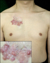
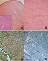
A 43-year-old Korean man developed multiple asymptomatic erythematous clustered plaques and nodules, and a skin-colored subcutaneous mass on the chest (Fig. 1). He had incurred a severe burn injury on the upper chest at the age of 8 years. The injury had healed with scar contracture but small nodules had repeatedly occurred; so, he excised the lesions 20 years ago. Although small nodules and plaques recurred several years later and spread gradually, he did not receive any checkup or treatment. Histological examination of the erythematous nodule and skin-colored subcutaneous mass revealed densely packed, monomorphous, plump spindle cells arranged in a storiform pattern. The cells extended into the subcutis, produced a characteristic honeycomb-like appearance, and were arranged in a fascicular pattern (Fig. 2A, B). Immunohistologic examination was positive for CD34 and negative for S-100 (Fig. 2C, D). These results were compatible with DFSP. The tumor was widely excised and, 6 months after the operation, there was no recurrence or distance metastasis.
DISCUSSION
The malignant transformation of burn scars constitutes a long-term complication of burns. Squamous cell carcinoma (SCC) is the most frequent burn scar neoplasm. Approximately 2% of burn scars undergo malignant transformation to SCC, while 0.3~0.5% undergo malignant transformation to basal cell carcinoma (BCC)3,4. Kowal-Vern and Criswell3 reviewed 412 cases of burn scar neoplasm from 146 articles published between 1923 and 2004. Of the tumors, 71% (n=293) were SCC, 12% (n=48) were BCC, 6% (n=23) were melanoma, 5% (n=21) were sarcoma, 4% (n=16) were other neoplasms, 1% (n=6) were squamo-basal cell carcinoma, and 1% (n=5) were squamous cell-melanoma3. Of the cases of sarcoma, only two were DFSP4,5. The pathophysiology of burn scar neoplasms is clearly distinct from that of regular skin cancer. Ewing's postulate requires: (1) evidence of a burn scar; (2) a tumor within the boundaries of the scar; (3) no previous tumor in that location; (4) tumor histology that is compatible with the cell types found in the skin and the scar; and (5) an adequate interval between the burn injury and tumor development6. The exact mechanism of malignant transformation in chronic wounds is unclear. However, some believe that constant irritation and infection, chronic ulceration, the release of local toxins following injury, poor lymphatic regeneration, repeated trauma, or exposure to noxious environmental agents can lead to the development of malignancies7,8. In our case, although chronic small nodules and plaques had recurred and spread, the patient did not undergo any treatment; therefore, constant, long-term irritation and local toxins may be to blame for the malignant transformation. Since sarcomas rarely arise in a burn scar, the clinical impression was SCC. Nevertheless, the histological findings of the lesion were compatible with DFSP. The tumor was widely excised and no adjuvant therapies were performed. Here, we report a very rare, third case of DFSP arising from a burn scar.
 XML Download