

 PDF
PDF ePub
ePub Citation
 Print
Print


INTRODUCTION
Granuloma annulare (GA) is a common, benign, inflammatory skin disease of unknown etiology that occurs in both adults and children1. Typically it is clinically characterized by papular lesions, usually distributed in ring-shaped configurations. Histologically the findings correspond to a necrobiotic granuloma, surrounded commonly by a radial arrangement of infiltrated lymphocytes and histiocytes2. The localized variant is the most common type and the acral sites are most commonly affected2,3. The ear is an unusual site for GA. Herein we report a rare case of bilateral GA on both ear anti-helical areas in a 28-year old man.
CASE REPORT
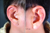
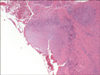
A healthy 28-year old man presented with multiple papules on both ear antihelical areas present for about 1 year. Physical examination revealed multiple, 1~5 mm, skin-colored, firm, non-tender papules on both ear antihelical areas (Fig. 1). The patient did not report subjective skin symptoms such as itching and pain. There was no previous history of trauma or chronic medical conditions, such as diabetes mellitus or rheumatoid arthritis. The patient was not routinely taking medication. The laboratory studies were within normal range except for the GPT (62 U/L); the tests included: a complete blood count, liver function, urine analysis and syphilis serology (VDRL). Excisional biopsy was performed of the right ear antihelical area. A biopsy specimen from the lesion of the right antihelical area showed a palisading infiltration of histiocytes, and lymphocytes around and between altered collagen fibers in the dermis (Fig. 2). Mucin was demonstrated within degenerative collagen bundles by the alcian blue stain (Fig. 3). After treatment with topical corticosteroid ointment and pimecrolimus cream (Elidel®, Novartis Pharma, Basel, Switzerland) for one month, the size of the lesions was slightly decreased.
DISCUSSION
GA is a benign, granulomatous disease that presents clinically as papules, nodules and plaques, often in an annular configuration. It is a self-limited disease, first described by Colcott-Fox in 1895 and Radcliffe-Croker in 19024. There are at least four clinical forms: (1) localized with single or multiple rings, (2) generalized, (3) subcutaneous, and (4) perforating2. Among the four clinical subtypes, the localized form is the most common type. It usually presents during the first three decades of life and has a female preference (2.25:1)3. About 50% of patients have solitary lesions. The most common affected areas are in the acral regions, especially the knuckles and dorsum of the fingers3. Facial involvement is rare3. Farrar et al.5 reported one case of perforating GA of both ears. In the Korean dermatology literature, there have been no reported cases of GA on both ears in the helical area.
The etiology of GA is unknown6. Some cases have been reported to follow an insect bite, trauma, sun exposure, tuberculin testing, PUVA therapy and viral infection including Ebstein-Barr, HIV, and herpes zoster4,6. In our case, there was no previous history of trauma, excess sun exposure, insect bites or medication use.
The histopathological features of localized GA include necrobiotic collagen and mucin in the dermis with surrounding inflammatory cells such as histiocytes, multinucleated giant cells, and a few acute inflammatory cells6,7. A palisading granuloma is the most characteristic finding of the histological lesion3. Considering the clinical and histological findings, the differential diagnosis of GA includes necrobiosis lipoidica, chondrodermatitis helicis nodularis and rheumatoid nodules3. Necrobiosis lipoidica shows dermal sclerosis and thickened subcutaneous septa and mucin deposits are uncommon. Chondrodermatitis helicis nodularis presents with painful nodules and has a characteristic layering of fibrin. Rheumatoid nodules histologically have fibrin rather than mucin deposits7.
GA has a spontaneous remission in about 50% of patients within two years. Recurrence occurs at the same site in about 40% of patients4,7-9. Reassurance and clinical observation may be the treatment of choice for localized, asymptomatic patients. Intralesion injections of triamcinolone and topical steroids are effective for individual lesions and are reasonable initial treatments8. Topical vitamin E, cryotherapy, intralesion interferon gamma, and surgical excision may be effective8. Other cutaneous treatment modalities include PUVA or UVA1 therapy, and CO2 laser treatment7. For generalized GA, many treatment modalities have been tried; this form is a chronic disease with a relapsing course that has shown a poor therapeutic response10,11. Systemic steroids may be very effective, but the side effects of high doses are dangerous. Dapsone, niacinamide, isotretinoin, sulfone, potassium iodide, chloroquine, alkylating agents, and electrodessication are used as additional options for treatment7,10. The patient in this case was treated with a topical steroid and a calcineurin inhibitor and the size of the lesions decreased slightly after one month of treatment.
This case illustrates a rare presentation of GA on the ear antihelixes with typical clinical and pathological findings.
 XML Download