

 PDF
PDF ePub
ePub Citation
 Print
Print


INTRODUCTION
Post-zoster eosinophilic dermatosis is one of the isotopic responses that occur in the region of healed herpes zoster. It was first described by Mitsuhashi and Kondo1 in 1997. The lesions in the first reported case clinically appeared as sharply demarcated brown plaques. Histologically, the lesions were characterized by a dermal infiltration of eosinophils and lymphocytes without atypicality. The dermatosis resolved one month after the application of topical steroid for 2 weeks1. To the best of our knowledge, only one case of post-zoster eosinophilic dermatosis, as an isotopic response, has been reported in the English medical literatures1. We report here on another case of post-zoster eosinophilic dermatosis in a 48-year-old woman with a history of herpes zoster infection.
CASE REPORT
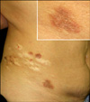
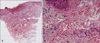
A 48-year-old female patient presented to our department with several severely pruritic, sharply demarcated, erythematous to brown papules and plaques with fine scales on her right flank (Fig. 1). The lesions had slowly developed for the past 6 months. The patient had no history of an insect bite, trauma or medication. Her medical history included herpes zoster along the T10 dermatome of the right flank, and this had had been successfully treated with an oral antiviral agent (Acyclovir, Vacrax®, 4,000 mg/day, 7 days) 13 years previously and she had residual scars. On laboratory examination, the complete blood cell count was within the normal limits. Eosinophils accounted for 3.8% (0~5%) of the peripheral white blood cells, and the total serum IgE level was 91.2 ku/L (<100 ku/L). The erythrocyte sedimentation rate was increased (35 mm/h). The skin biopsy specimen obtained from a plaque on the right flank revealed mild acanthosis and spongiosis in the epidermis and a perivascular inflammatory cell infiltration composed of abundant eosinophils, lymphocytes and histiocytes in the dermis (Fig. 2). There was no granulomatous change. An application of a topical steroid (Dethasone ointment®, bid) for 2 weeks resulted in the lesions clearing up; no sign of recurrence was noted 6 months after treatment.
DISCUSSION
Wolf et al.2 define an isotopic response as the occurrence of a new skin disorder at the site of another unrelated and already healed skin disease. The first disease in most of these cases is herpes zoster, and several cutaneous reactions have been described in the dermatomes affected by herpes zoster3. The second diseases may be granulomatous reactions (mainly granuloma annulare), malignancies (single tumor and leukemic or lymphomatous infiltration), immune disorders (e.g. lichen planus, allergic contact dermatitis), infections (viral, bacterial, fungal) and others (e.g. acneiform lesions, reactive perforating collagenosis)3-5. Seven cases of postherpetic isotopic reactions have currently been reported in the Korean dermatologic literature (Table 1)6-13.
The pathogenesis of isotopic responses is unclear. Several possibilities have been considered: a viral origin, an immunologic origin, a vascular origin and a neural origin. Concerning a viral origin, it has been suggested that either the virus is directly responsible for the second disease, or the second disease is the result of an inflammatory reaction that develops in response to residual viral antigens or to the tissue damage. Polymerase chain reaction has been used to investigate the skin lesions for the presence of varicella-zoster virus, which has been consistently found only in the early lesions3. The development of post-zoster dermatosis may be influenced by long-lasting immunologic changes, alterations in the microcirculation or the destruction of A δ and C nerve fibers that occurs after viral infection of the skin4,14.
Our patient presented with several erythematous to brown papules and plaques with fine scales on the T10 dermatome in the area of a previous zoster infection. The clinical differential diagnosis in this case included granuloma annulare and lichen planus. However, the absence of any histological granulomatous change ruled out the possibility of granuloma annulare. Lichen planus was also excluded because of the absence of the characteristic histological findings such as basal layer vacuolization, Civatte bodies and a band-like inflammatory cell infiltration. Instead, one of the most pronounced histological features in our case was a prominent eosinophil infiltration in the lesion. Thus, we needed to further exclude the possibility of drug eruption and insect bites, but the patient denied any medication history or insect bite history, and peripheral eosinophilia was not detected. Based on these clinical and histopathological findings, we diagnosed our case as post-zoster eosinophilic dermatosis. Post-zoster eosinophilic dermatosis is one of the isotopic responses, and these are clinically and histopathologically distinct from the other known dermatoses. It occurs on the region of the healed herpes zoster and it is histologically characterized by a dermal infiltration of eosinophils.
In an isotopic response, the time elapsing between the first disease and the second one widely varies, ranging from days to years1-4. In our patient, the delay was approximately 13 years. Until we better understand the pathogenesis of the isotopic response, it will be difficult to design targeted therapies3,7. Langenberg et al.15 suggest that antiviral therapy may not be appropriate in inflammatory post-zoster conditions. Instead, topical or systemic corticosteroids are often used7,15,16. Most post-zoster conditions are self-limited in nature, but these lesions should be examined histologically because of the possibility of occurrence of some malignancies such as lymphoma and Kaposi's sarcoma16,17.

 XML Download