

 PDF
PDF ePub
ePub Citation
 Print
Print


INTRODUCTION
Keloids are benign, hard and persistent fibrous proliferations that develop in predisposed persons at sites of cutaneous injury1-3. Keloids of the ear usually appear as shiny, smooth and globular growths on one or both sides of the ear. They are usually preceded by ear piercing, trauma or burns1,2. The incidence of keloid formation is difficult to assess as it varies from 4.5% to 16% in the Black and Hispanic populations4,5. The incidence of keloid in dark-skinned people is estimated to be 3 to 20 times that of light-skinned people1,5-7.
In some cases, nonsurgical therapies such as intralesional corticosteroids8, X-ray irradiation9, pressure dressing10, silicone gel sheets11, cryotherapy12 and interferon13 were reported to be effective to treat keloids. However, even though the size of the keloid may decrease with nonsurgical treatment alone for the cases of larger lesions, the results may not be cosmetically satisfactory. Various surgical methods such as excision followed by primary suture14, healing by secondary intention15, skin graft16 or local flap17 have been used. Yet surgical excision alone has shown varying degrees of success and perioperative nonsurgical therapies should be combined to prevent the inevitable recurrence after surgical excision.
The purpose of this study was to study the usefulness of a combination of surgical excision and perioperative corticosteroid intralesional injection, which is a very economical treatment that patients will comply with, for the treatment of earlobe keloids in an Asian population.
MATERIALS AND METHODS
Patients
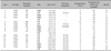
From 1997 to 2006, eighteen earlobe keloids in fifteen Korean patients were treated with our protocol. The patients' ages ranged from 15 to 32 years (mean age: 24 years). All the patients were female and the keloids occurred after ear piercing. At the time of treatment the lesions have been present from 8 to 60 months (mean: 28.7 months). Seven patients had keloids on the left ear and five patients had keloids on the right ear. Three patients had bilateral lesions. The size of the keloids ranged from 0.25 to 3 cm2 (mean: 1.25 cm2). Among the fifteen patients, eight patients had received prior treatments with intralesional corticosteroid injection and one patient had received excision (Table 1).
Methods
Preoperative intralesional triamcinolone acetonide (TA) injections were administered twice at a 1-month interval. A total of 0.1 to 1.0 ml of TA (20~40 mg/ml) was decided upon depending on the size and the activity of the lesions. The surgery was performed under local anesthesia with using 1% lidocaine and 1 : 100,000 epinephrine. An incision was done within the edge of the keloid. The marginal skin of the overlying keloid, which consisted of epidermis and the thin dermis overlying the keloid, was raised from the fibrous keloid core. The fibrous keloid core was completely removed after completely raising the marginal skin. After trimming the redundant marginal skin, the defects were resurfaced using the marginal skin of the overlying keloid. All the wounds were closed with using one layer of 6-0 Ethilon without tension. A simple dressing was applied after surgery. The postoperative intralesional TA injection was started at a 2 week time point after surgery and an injection was given every 1 month for several months, depending on the patient's clinical progress, and a total of 0.1 to 1.0 ml of TA (20~40 mg/ml) was also decided upon depending on the size and the activity of the remaining lesions.
The final results were judged during the postoperative follow-up period. A good result was defined as a normalappearing, flat scar that needed no further aesthetic refinement. A fair result was defined as a flat scar without recurrence, but aesthetic refinement was needed. Recurrence was defined as any indurated papule or nodule extending beyond the borders of the healed scar line of the previously excised lesion.
RESULTS
The follow-up period ranged from 4 to 42 months (mean: 18.5 months). After the surgery, TA intralesional injection was performed from 2 to 13 times (mean: 5.2 times).
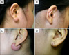
Among the treated keloids, 11 showed good results (Fig. 1), 4 showed fair results and 3 recurred (Table 1). We performed dermabrasion on the lesions with fair results. For case 6, an earlobe keloid recurred about 4 months after the surgery. Preoperative intralesional TA injection was administered twice at a 1-month interval and postoperative intralesional TA injections were done 4 times every 1 month. Yet the lesion enlarged during the course of performing the postoperative intralesional TA injections. After the recurrence, the patient did not revisit our clinic and we could not provide further treatment for her recurred earlobe keloid. For case 15, both earlobe keloids recurred about 8 months after surgery. She wanted to be transferred to another clinic due to personal reasons and we recommended regular intralesional TA injections every 1 month. For cases 10 and 12, indurated papules on the healed scar line occurred 1 month after the surgery. We treated the indurated papules with intralesional TA injection twice at a 1-month interval and the indurations were resolved. Any adverse effects such as atrophy and telangiectasia were not observed at and around the injection sites.
DISCUSSION
Keloid development has been associated with different types of skin injury, including surgery, ear piercing, laceration, burn, vaccination or inflammatory processes1,2. With an ever-expanding number of patients undergoing body piercing, dermatologists are increasingly being consulted for the treatment of ear keloids. Multiple therapeutic options are available for the treatment of keloids. Combination therapy appears to be the most effective, although few previous studies have compared the various regimens.
Excision alone of keloids has been associated with recurrence rates of 45% to 100%18,19. Numerous treatments after excision have been used such as postexcisional injections of corticosteroids8 or interferon13, radiotherapy9, pressure splints10 and silicone sheeting11. In a large review of the literature, Friedman20 proposed surgical excision and corticosteroids for small keloids, pressure therapy and a combination of surgery and steroid for moderately large scars and a combination of surgery and postoperative radiotherapy for very large, resistant scars. Earlier studies have shown that postexcisional TA injection is associated with a recurrence rate of 0% to 100%, but the majority of studies reported less than a 50% recurrence rate18. The recurrence rate in our study was 16.6%.
Berman and Flores13 reported a recurrence rate of 18.7% when interferon alpha-2b injections were given after keloid excisions. Their result was lower than the recurrence rate achieved with excision alone (51%) or a combination of excision and postoperative triamcinolone intralesional injection (58%). Although the recurrence rate of interferon injection after excision is known to be low, interferon treatment is more expensive than TA intralesional injection and the results are similar to ours.
Recurrence rates of as low as 2%, but up to 50%, have been reported following postoperative external-beam radiotherapy21,22. A randomized controlled trial of earlobe keloid excision followed by either intralesional steroid injection or radiotherapy has shown that radiotherapy has a lower recurrence rate than that of steroid injection, a statistically significant difference was not observed23. Although surgical excision combined with radiotherapy has shown fairly successful results in treating resistant keloids, many surgeons are still reluctant to use ionizing radiation for the treatment of benign diseases.
Surgery followed by pressure treatment has shown a good response rate of 90~100%, and especially after excision of earlobe keloids10. However, the pressure must be maintained day and night for a minimum of 6~9 months, and premature release is frequently followed by recurrence of the lesion.
Topical silicone sheets have been shown to soften scars and reduce itching, and children like this treatment because the gel-sheeting is painless. It usually takes 6~12 months of therapy to achieve the best results, but most patients become noncompliant after several months because of the treatment's duration and the inconvenience of cutting and placing the silicone gel-sheeting on the keloid11.
The postsurgical use of imiquimod 5% cream has recently been reported24. All 4 patients were successfully treated without recurrence for 12 months after their final excision. Although imiquimod 5% cream following tangential shave excision was effective, most patients experienced mild to marked irritation secondary to the daily application. Those with marked irritation will sometimes have to discontinue the medication for several days to a week. The patients who have large surgical sites and wounds closed with flaps, grafts or tension should not start imiquimod cream therapy for four to six weeks postoperatively because early application often causes the surgical site to splay or dehisce.
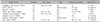
The use of steroid injections as an adjunctive procedure after keloid excision had been reported with using various dosages, schedules and concentrations of drug. When excision was combined with postoperative intralesional triamcinolone acetonide injection, the recurrence rates were 0~86.7% (Table 2)8,13,23,25-28. Table 2 showed that the recurrence rates (0~33.3%) for the ear keloids were lower than those for keloids other sites and our study showed a 16.6% recurrence rate. Shons and Press8 reported that only 3% of earlobe keloids recurred after excision and three injections of 40 mg/ml triamcinolone at a 4 week interval and beginning 3 weeks postoperatively. Singleton and Gross25 reported on the treatment of auricular and lobular keloids using a posterior approach for excision followed by steroid injection at immediately postoperative period. Steroid injections were then continued at a 1-month interval postoperatively for 1 year. For the 21 patients who were followed for more than one year, there were 13 good results and 2 recurrences. For the 33 patients who were followed for less than one year, 24 showed good results and 2 recurred. The corticosteroid inhibits alpha2-macroglobulin, which in turn inhibits collagenase. Once this pathway is blocked, collagenase is elaborated, thus enabling collagen degeneration29.
The risk of relapse following these treatments depends on local wound-related factors and general patient-related factors. A high recurrence rate is seen at sites where have keloids developed after tensioned wound closure, wound infection and dehiscence. Patients with a positive family history and a past history of recalcitrant keloids with prior treatment failures are at a greater risk of recurrence30.
The high rate of recurrence following excision alone has led to the investigation of many different types of adjuvant therapy, but of these techniques has been demonstrated to be optimal. Until one strategy is shown to be clearly superior, the choice of adjuvant treatment needs to be tailored to the individual patient and the clinical situation. Although various treatments after surgery have previously been used, we think that excision with corticosteroid intralesional injections can be used as the standard treatment for earlobe keloids.
 XML Download