

 PDF
PDF ePub
ePub Citation
 Print
Print


INTRODUCTION
Membranous lipodystrophy represents a peculiar type of fat necrosis that is present in patients with diverse kinds of skin disease. Nasu et al1, in 1973, described it as a clinical entity with cyst-like lesions of fat occurring in the long bones accompanied by sudanophilic leukoencephalopathy. Histologically, at the border of the lobules with the septa, there are fat cysts lined by a thin, homogenous, eosinophilic layer of protein with fine, feathery projections extending into the fat cavity. These particular changes in the fat tissue have been shown to be related to many local and systemic diseases, including lupus erythematosus, diabetes mellitus, erythema nodosum, morphea, trauma, atypical mycobacterial infection, sclerosing panniculitis, and vascular disorders. Still, in some cases, no underlying disease is found.
Lichen amyloidosis (LA) is a form of primary, localized, cutaneous amyloidosis in which amyloid is deposited in previously normal skin without any evidence of visceral involvement. Its etiology remains unknown, although it has a genetic predisposition2. Clinically, small, discrete, pruritic, hyperkeratotic, hyperpigmented papules are noted, mostly on the extensor surfaces of the lower extremities.
We report a case of subcutaneous membranous lipodystrophy in a patient with lichen amyloidosis.
CASE REPORT
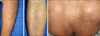
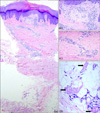
A 72-year-old man presented with multiple, dark brownish, hyperkeratotic papules on the arms, legs, and back (Fig. 1), which had been present for the last 8 months. These lesions were severely pruritic. The patient had noted gradual progression of the lesions, especially in recent times. His medical history was significant for a cerebral infarction occurring 10 years prior to presentation. On physical examination, the patient had neither musculoskeletal nor mental abnormalities. Laboratory studies, including complete blood cell count, routine chemistry profile, and urinalysis, were within normal limits. The chest radiograph was normal. A skin biopsy specimen was taken from a papule on the right shin. The biopsy specimen was stained with hematoxylin-eosin. Histopathologic examination revealed hyperkeratosis and irregular acanthosis of the epidermis (Fig. 2A) and homogenous, eosinophilic material in the papillary dermis (Fig. 2B). The blood vessels in the dermis showed thickened walls, dilatation, and proliferation and were surrounded by inflammatory cells (Fig. 2C). In the subcutaneous fat layer, inflammatory cell infiltrates were seen, as were eosinophilic, feathery lipomembrane projections into the fat lobules (Fig. 2D). On the basis of these clinical and histological findings, a diagnosis of membranous lipodystrophy with lichen amyloidosis was made. Oral anti-histamines and a topical corticosteroid were prescribed, and the patient's pruritus gradually decreased. Now we are observing the progress of the disease.
DISCUSSION
The term "amyloid" (starch-like) was neologized in 1854 by Virchow, who noted its resemblance to starch or cellulose. Rokitansky first described amyloidosis in 18422. Lichen amyloidosis is a primary cutaneous disease in which amyloid material is deposited in previously normal skin without evidence of visceral involvement. Characteristically, the lesions are made up of extremely pruritic, small (2~3 mm in diameter), discrete, waxy brownish papules, which are conic or hemispheric in form and firm to hard in consistency. Papules usually appear in groups on the extensor surfaces of the extremities, particularly the legs. The etiology of lichen amyloidosis remains unknown, but chronic irritation of the skin has been suggested as an etiological factor3. Recurrent external physical irritation such as repeated friction or other trauma, medication, and solar injury have been suspected as precipitating factors. Histologically, amyloid deposits are mostly limited to the upper dermis and arise because of focal epidermal damage with subsequent conversion of necrotic keratinocytes into amyloid in the papillary dermis.
Membranocystic fat necrosis is a unique alteration in adipose tissue. Nasu et al1 first described the disease in 1973 as a genetic disorder characterized by profound membranocystic degeneration of long bones and systemic adipose tissues, associated with progressive sudanophilic leukodystrophy of the brain. These particular pathologic findings have been observed in various clinical conditions other than those mentioned above, including arterial ischemia, venous insufficiency, and other dermatologic diseases. Therefore, membranous lipodystrophy is divided into two categories-primary and secondary membranous lipodystrophy-based on the presence of underlying disease.
The etiology of membranous lipodystrophy is unknown. Ahn et al4 proposed seven hypothetical pathologic mechanisms: 1) idiopathic processes; 2) enormous proliferation of fat cell membranes; 3) physiochemical interaction between the ground substance in connective tissue and fat droplets; 4) free fatty acids released from degenerated fat cells processed by macrophages to produce membranous lipodystrophy; 5) a metabolic disorder of lipids in mesenchymal cells; 6) loops and folds in the basal laminae of fat cells in varying stages of lipid depletion at the time of necrosis; 7) ischemic injury of adipose tissue resulting from venous insufficiency. Lee et al5 reported two cases of membranous lipodystrophy-like changes in traumatic lipogranulomas caused by safety belts. Poppiti et al6 proposed trauma as the one of the possible causes of membranous lipodystrophy.
On histopathological examination, variably sized cysts are surrounded by anuclear, eosinophilic membranes in the subcutaneous fat layer. These membranes often project into fat lobules, creating a pseudopapilla or arabesque appearance. The eosinophilic membranes are considered to be ceroid. Ceroid is an oxidant of unsaturated fatty acids produced in metabolic disorders, released as a result of lipocyte necrosis, and phagocytized by macrophages7.
The current patient had lichen amyloidosis, as well as membranous lipodystrophy. Severe and intractable pruritus led to repetitive scratching, which exacerbated trauma to the skin. As mentioned above, repeated trauma has been proposed as one of the etiologic factor in membranous lipodystrophy. Our histopathological examination revealed dermal vascular proliferation, dilatation, and perivascular inflammatory cellular infiltration. These findings might indicate vascular insufficiency or damage, which are considered the most important factors in membranous lipodystrophy. Alegre et al8 reported 13 patients with lipomembranous changes in the subcutaneous adipose tissue. In more than half the patients, histopathologic evidence of endarteritis obliterans, venous stasis, and hemorrhage was present, suggesting that membranocystic changes may be the result of ischemic injury to the adipose tissue.
The relationship between lichen amyloidosis and membranous lipodystrophy is not established. But the features of this case suggest that lichen amyloidosis might give rise to membranous lipodystrophy based on the mechanisms mentioned earlier.
Membranous lipodystrophy accompanies various dermatologic disorders, such as morphea, lupus erythematosus, erythema nodosum, stasis dermatitis, and steroid panniculitis9. However, there has been no report of membranous lipodystrophy accompanying lichen amyloidosis. We reported a case of lichen amyloidosis in which membranous lipodystrophy was found in the subcutaneous fat tissue.
 XML Download