

 PDF
PDF ePub
ePub Citation
 Print
Print


INTRODUCTION
Hypertrichosis is the excessive growth of hair on the non-androgen dependent areas of the body. Primary hypertrichosis has been classified based on the age of onset as to whether it is congenital or acquired, and the extent of distribution takes on either localized or generalized forms. Primary localized hypertrichosis may occur as hypertrichosis cubiti involving the elbows, anterior cervical hypertrichosis, posterior cervical hypertrichosis or a faun tail deformity1. A faun tail is a lock of coarse, terminal hair situated on the lumbosacral area. According to the Korean dermatologic articles2-5, only four cases with faun tail have been reported in association with tethered cord syndrome or spina bifida occulta. We report herein on an interesting case of a bazaar faun tail naevus on a 36-year-old male.
CASE REPORT
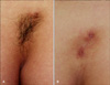
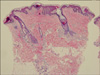
A 36-year-old man was referred to our dermatology outpatient clinic for the treatment of a congenital focal hypertrichotic area on the lumbosacral region because it was cosmetically embarrassing. The dermatologic examination revealed a localized, 3.0×5.0 cm sized, reverse triangular shaped hair tuft on the lumbosacral region. Coarse, dark, terminal hairs were observed and there was a single skin colored protruding papule within the hairs (Fig. 1A). The lesion had been present since his birth. The past medical history was noncontributory except for psoriasis vulgaris. No motor weakness or sensory changes were observed on the neurological examination. His height was estimated to be 156 cm. There was no history of a similar lesion in the family and relatives. The complete blood cell count, erythrocyte sedimentation rate and blood chemistry were all within the normal ranges. Simple lumbosacral radiography showed no specific finding. The magnetic resonance imaging scan showed no specific finding, except for a left subarticular protrusion of L4-5 and the loss of lordosis of the lumbar spine. The skin biopsy from the hypertrichotic patch demonstrated normal epidermis and a superficial perivascular lymphocytic infiltration with mature terminal hair follicles in the dermis (Fig. 2). The other biopsy revealed a skin tag. A diagnosis of faun tail without any underlying neurologic disease was made. He was periodically treated with intense pulsed light (IPL) for hair removal and he was well satisfied with the result (Fig. 1B).
DISCUSSION
Hypertrichosis is a condition of excessive hair growth that must be distinguished from hirsutism, which is characterized by an androgen-dependent hair pattern with an excessive body and facial terminal hair distribution in a male pattern1. Localized hypertrichosis can be categorized into the acquired and congenital forms. Congenital localized hypertrichosis is present at birth or early in life without any inducing factors1. Congenital localized hypertrichosis is most commonly located in the sacral area and this is called "faun tail".
The mechanisms for excess hair growth have not been well established. Clinical evidence exists that localized or site-specific factors may be important in determining the fate of a hair follicle. Conversion of vellus or fine caliber hairs to thicker terminal hairs occurs as a result of hormonal changes, but only at androgendependent sites such as the beard, chest, axillae and groin6. Congenital localized hypertrichosis in hamartomas or underlying neurologic abnormalities may represent an alternative pathway. Recent research has suggested the tremendous plasticity of the hair follicle, its mesenchyme and the surrounding connective tissue sheath, which allows for dramatic transformations of hair follicles Localized hypertrichosis may therefore be a reflection of an abnormal signal for enlargement of the follicular papilla, a prolongation of the normal anagen hair growth cycle or both7.
The clinical importance of a midline cutaneous posterior anomaly is that they are frequently associated with underlying defects such as diastematomyelia, meningocele, spina bifida, kyphoscoliosis or chest deformities. As both skin and nervous tissue are of an ectodermal origin, anomalies of these tissues may occur simultaneously. When the congenital localized hypertrichosis is located away from the spine in apparently normal skin, it has been named as simple nevoid hypertrichosis, and it is usually a totally benign lesion8. The cutaneous lesions that should raise a higher degree of suspicion include hypertrichosis, dimples, aplasia cutis, lipoma, hemangioma, dermoid cyst or sinus, acrochordons, true tail, pseudotail and congenital scarring9,10. Thus, localized hypertrichosis may be a clue for an underlying defect, some of which are surgically correctable at an early age. Sacral hypertrichosis is also known as faun tail naevus, and this is the most common skin lesion that is evident at birth11, as was seen in our patient. Hairy patches are most frequently associated with tethered cord and diastematomyelia12. Although our case demonstrated no underlying neurologic problem associated with his localized hypertrichosis, accurate screening modalities such as MRI are necessary to detect any underlying dysraphic anomalies and to prevent the occurrence or progression of the neurologic deficit by timely intervention before neurological damage has occurred.
As for the management of hypertrichosis, long-term removal of unwanted hair is a challenge. The need for treatment depends on the degree of hypertrichosis and the psychosocial needs of the patient. The currently available treatment methods for removal of excessive hair include bleaching, trimming, waxing, physical and chemical depilatories, electrolysis, intense pulsed light therapy and laser hair removal13. A variety of laser devices and intense pulsed light therapy are now available. All are based on the principle of selective photothermolysis; the melanin pigment in the hair follicle provides the chromophore for selective targeting of hair follicles, while the surrounding dermis is spared. Therefore, at deeply penetrating wavelengths in the 600~1,100 nm range, melanin absorption may be used for selective photothermolysis of hair follicles14. Intense pulsed light source (590~1200 nm) therapy for hair removal seems to be more effective for darker hair15. Because our patient's lumbosacral hair was dark and thick like his axillary hair, we choose intense pulsed light therapy for hair removal. Occasionally, posttreatment erythema, edema, blisters and hyperpigmentation can occur.
The aim of this paper is to draw attention to faun tail and to emphasize the responsibility of physicians and cosmeticians to the recognize early in patients' lives lumbar paraspinal skin lesions so that appropriate early treatment can be administered for the possible underlying anomalies before the irreversible sequelae develop. Although otherwise benign, these disorders may result in cosmetic disfigurement and psychosocial trauma for the patients. These patients should be adequately advised of the available treatment methods for both temporary and permanent hair removal.
 XML Download