

 PDF
PDF ePub
ePub Citation
 Print
Print


INTRODUCTION
Congenital melanocytic nevus (CMN) is defined as a tissue malformation of the neuroectoderm that presents at birth or it appears within the first few months of life1,2. CMNs are usually classified according to their current size or the size that the CMN is predicted to attain at adulthood: small (less than 1.5 cm in diameter), medium (1.5~20 cm) and large (more than 20 cm)3. The classification system is important because it has an effect on the management of CMNs. In addition to size, several factors that are considered for the clinical management of CMN include the location, thickness, clinical appearance, the risk for developing melanoma, the psychological effect and the cosmetic component4. Thus, the management of CMNs needs to be individualized for each patient5. There are many treatment options for CMNs: surgical excision, dermabrasions, curettage, laser treatment, chemical peels and cryosurgery1,6. Surgical excision is the most commonly used method because this completely removes the nevus cells. However, it requires two or more staged procedures in many cases, and physicians should consider the complications according to surgery, such as a postoperative long linear scar. In this background, lasers are now being increasingly used for treating large lesions and the CMNs located on cosmetically sensitive areas. But lasers are effective only for removing nevus cells of the upper dermis and they can not remove the deeper portion of the nevus cells. In addition, laser therapy requires multiple treatment sessions. Therefore, we introduce here a new method to treat medium-sized CMNs with the combination of surgical excision and Er:YAG laser ablation.
MATERIALS AND METHODS
Patients
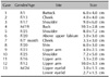
Since 2004, we have treated 13 patients using a combination of surgical excision and Er:YAG laser ablation. Informed consent was obtained from all the patients. The patients were two males and eleven females aged 7 months to 25 years. The CMN was located on the extremities in seven subjects, on the trunk in two subjects and on the face in four subjects (Table 1). All the patients had Fitzpatrick's skin types III and IV.
Methods
First, we excised the nevus as much as possible, including the deeply pigmented or elevated portion, and especially the hair-bearing area, after administering local anesthesia (2% lidocaine with 1:80,000 epinephrine). Various designs of excision, including an elliptical shape, were used according to the size and location of the nevus. Second, only dermal suturing using vircryl was performed after the surgical dissection for achieving tension relief of the skin without suturing the epidermis. Third, we ablated the residual pigmented areas, including the suture lines, with using a dual-mode 2,940 nm Er:YAG laser (Contour, Sciton, Inc., Palo Alto, CA, USA). On the average, three to five passes of ablation were done in 90~100 µm ablation mode and using a 4 mm laser beam spot at 25.0 J/cm2. Ablation was performed until the visible pigmented part was diminished and an even wound surface was created. After the laser treatment, the wound was dressed with a chlorhexidine paraffin gauze-based dressing material (Bactigras, Smith & Nephew Co., Hull, UK) and topical antibiotic ointment. The occlusive dressing was changed with using hydrocolloid dressing material at an interval of three days for two weeks until the re-epithelialization was completed.
Follow-up
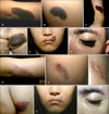
All the lesions were followed up for 6 months. At each visit, the treated areas were evaluated with respect to the healing status, infection, postoperative hyperpigmentation and hypopigmentation, erythema, scarring and repigmentation. Photographs were taken prior to treatment and at each visit by using a Canon EOS 300 D digital camera (Canon Inc., Tokyo, Japan). The Global Assessment Scale (GAS) scores were checked to assess the overall results and the degree of the patient and physician satisfaction with the therapy at the 16th week according to the following scale: excellent, good, fair and poor. The postoperative changes, including erythema, pigmentary change and hypertrophic scarring, were also estimated by the physicians who were involved in the treatment; the postoperative changes were rated according to the following scale: none, mild, moderate and severe.
RESULTS
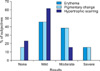
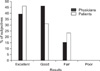
The treatment was well tolerated by all the patients and no major complications occurred. Re-epithelialization was complete within 10~15 days in most cases. All the patients showed good to excellent results for a one-stage procedure (Fig. 1). With respect to the investigators' GAS scores, five patients (37%) were rated as excellent and six patients (46%) were rated as good. Especially for the cosmetic outcome, this combination method showed excellent results and this was confirmed by the subjects' GAS scores, which were checked at the 16th week follow up exam (Fig. 2). For the GAS scores by the patients, six patients (46%) were rated as excellent and four patients (31%) were rated as good. Infections did not occur in any of the patients. Erythema was observed in all patients, but this was rated as mild to moderate in 11 (83.6%) of 13 lesions and it gradually subsided. Pigmentary changes, including postoperative hyperpigmentation, hypopigmentation and repigmentation, were observed in 11 patients, and six patients (46.2%) showed mild changes and three patients (23.0%) showed moderate changes. Two patients showed a severe degree of repigmentation and we further treated these lesions with pigment-specific Q-switched Nd:YAG lasers (Medlite IV, Medlite, USA). Then the pigmentation became decreased. Three patients showed no signs of hypertrophic scarring, but most of the patients (61.6%) showed a mild degree of hypertrophic changes (Fig. 3). All the lesions were followed up for 6 months. Although our cases showed excellent clinical results, long-term follow up will be needed for assess the lesions for recurrence of the nevus or delayed scarring.
DISCUSSION
The management of CMN is a challenge to physicians because there are several factors to consider for deciding on the proper treatment modality, including the patient's age and the lesion's size, location, depth, shape and malignant potential. But no specific guidelines have been established for treating CMNs5. There are many treatment options for CMNs: surgical excision, dermabrasions, curettage, laser treatment, chemical peels and cryosurgery1,6,7. To decide on the modality of treatment, we should consider two main issues8. One is the potential for malignant transformation3,9,10 and the other is the cosmetic component, and particularly for large-sized CMNs or those CMNs located on cosmetically complex areas, including the face and genital area6,11. Surgical excision is the most commonly used method and it requires full reconstructive methods such as direct excision with primary closure, including serial excision, skin grafting, tissue expansion, free tissue transfer etc1,12,13. Complete and full-thickness excision is an ideal method in terms of preventing the development of melanoma and for cosmetic reasons. But in most cases, except for the small CMNs, serial excision or tissue expansion will be needed so that multiple procedures are required for complete removal. Also, creating a long linear scar and scar widening due to tension should be considered and there are many complications after tissue expansion, including deflation or disruption of the expanders, hematoma, severe infection and flap ischemia1,5,12. On the other hand, laser treatment is useful for large-sized CMNs, it is effective for cosmetically sensitive areas and it is safe for children14. The lasers used for the treatment of CMN include pigment-specific lasers15-19, resurfacing lasers20-23 and a combination of the two14,17,24,25. In particular, the Er:YAG laser is precise and it can be used for controlled resurfacing. It causes significantly less deep thermal injury, there is a shorter re-epithelialization time and it is less painful compared with the CO2 laser20,23. But it is only effective with respect to the upper levels of the dermis and multiple treatments at variable intervals can be required for achieving a cosmetically satisfactory outcome.
Therefore, we introduce a new method to treat medium-sized CMNs with using the combination of surgical excision along with Er:YAG laser ablation. This method has some advantages over other modalities. First, it enables a much larger volume of lesion to be removed in a one stage procedure. It is the time-saving method and it can promote early recovery. Second, it can remove most of the deep dermal components by excision and this had been confirmed to prevent the development of melanoma, as compared with laser treatment only. Third, the suture line with this combined technique can be shorter as compared with radical excision of the whole nevus and it does not sacrifice the normal adjacent skin. Moreover, the formation of a surgical scar can be diminished because of the laser ablation over the suture line. Fourth, it is beneficial for treating CMNs located on cosmetically sensitive areas. There were no severe complications associated with the procedure, and only mild erythema and pigmentary change were observed. Particularly, the patients treated by this method reported high satisfaction scores. Ten (77%) of the total 13 patients in our study reported the cosmetic results to be good to excellent at four months after treatment.
We report here that the combination of surgical excision and Er:YAG laser resurfacing is a safe, effective method for managing medium-sized CMNs because it is an one-stage procedure that causes less scarring. Further, this combined treatment showed a good cosmetic outcome with a high degree of satisfaction, as reported by both the physicians and the patients. We suggest that this combined treatment will be a good option for treating medium-sized CMNs.
 XML Download